Astrocytoma
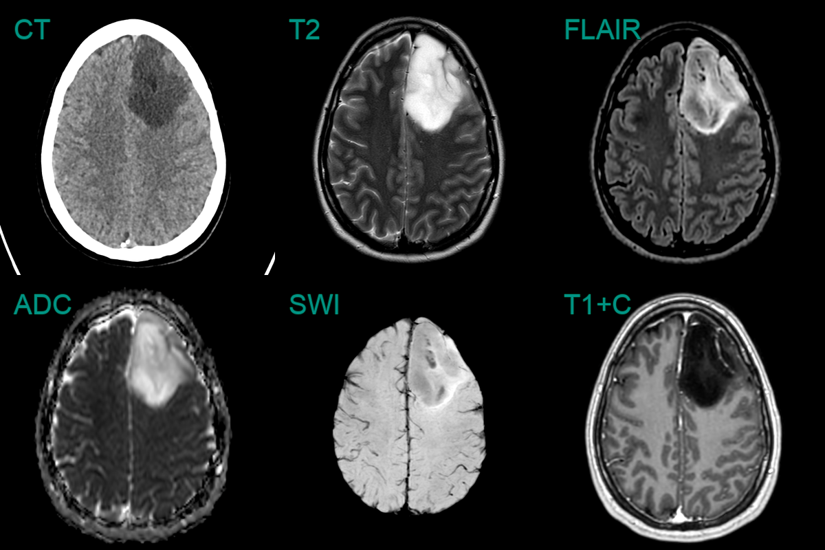
- 35-year-old patient with 2 month history of headache presented after a tonic-clonic seizure.
- Imaging showed a quite well defined T2-hyperintense non-enhancing lesion.
- Low FLAIR signal in more than half of the tumour representing the T2-FLAIR mismatch sign suggested an astrocytoma that was confirmed on histopathology following resection.
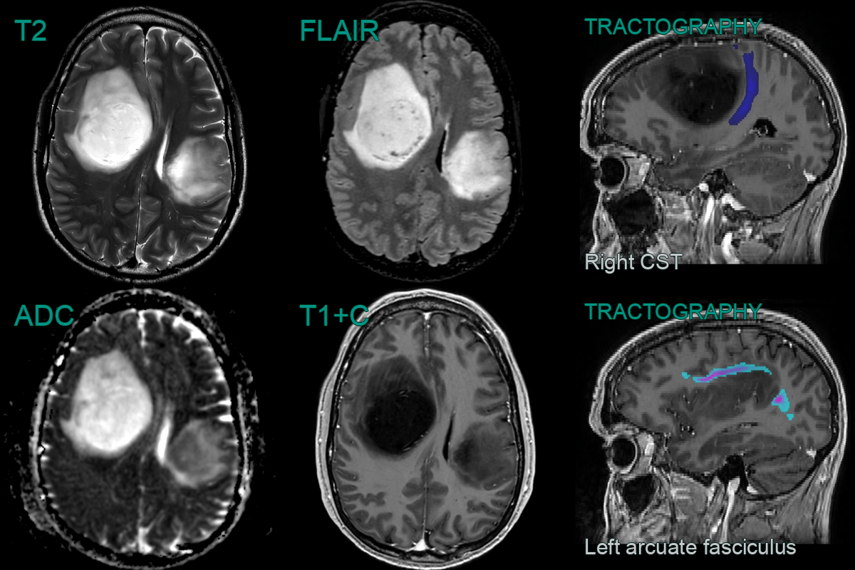
- A 40 year old presented with with memory issues and headache.
- MRI showed two large lesions, one in each hemisphere that did not show any enhancement but there were low ADC values within the left sided lesion.
- Initial histopathlogy suggested a grade 2 astrocytoma however, following molecular anlysis, this was upgrade to a grade 4 astrocytoma on the basis of a CDKN2A/B deletion.
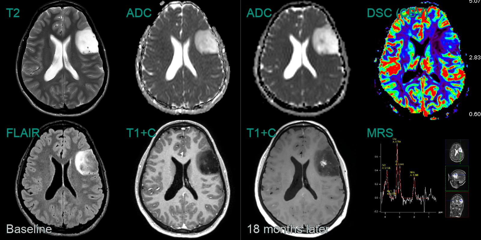
- A 20-year-old patient with Li Fraumeni Syndrome presented following a seizure.
- MRI showed a left frontal lesion with a T2-FLAIR mismatch (hyperintense on T2 and more than half of the lesion hypointense on FLAIR).
- On follow-up imaging 18 months later, spiculated enhancement developed iwthin the tumour, which corresponded to an a region of low values on ADC.
- CBV was elevated (ratio of 4 relative to normal appearing brain tissue) and MR spectrscopy showed reversal of Hunter's angle (elevated choline and reduced NAA).
- Following resection, a grade 3 astrocytoma was diagnosed.
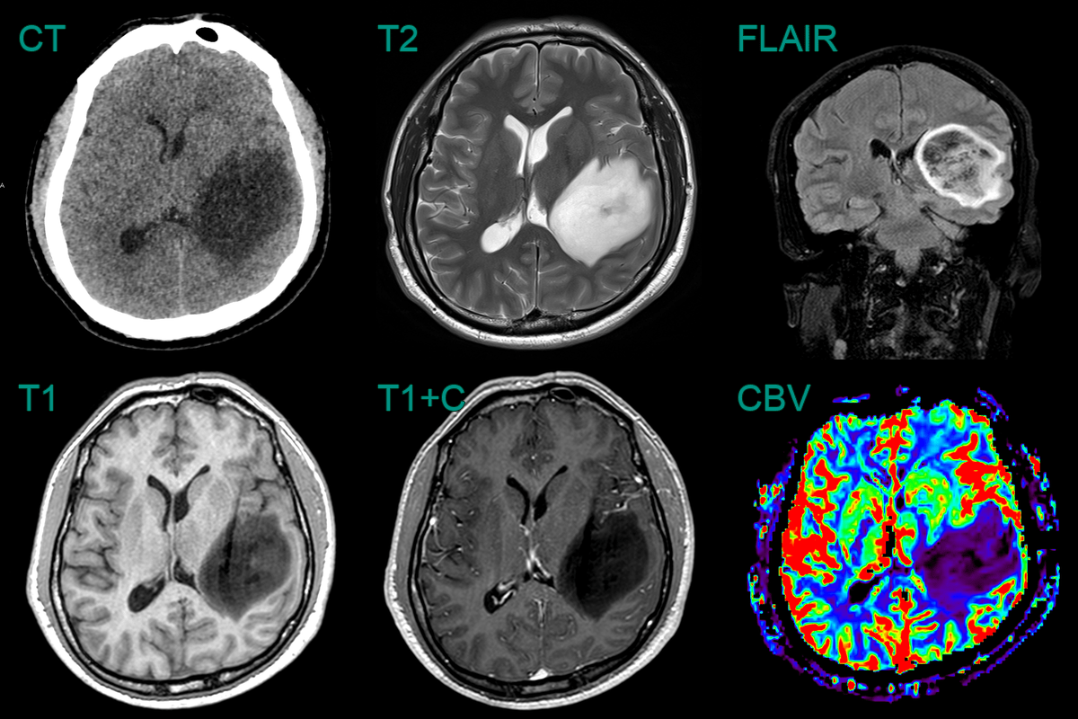
- A 30-year-old patient presented following a seizure.
- MRI showed a large right parietal lesion with a T2-FLAIR mismatch.
- A region of enhancement corresponded to a region of relative hypercelluarity and higher CBV.
- MRS was very abnormal with grossly reduced NAA, elevated choline, and the presence of lactate.
- Final molecular diagnosis was a grade 4 astrocytoma.


