Cerebral Autosomal Dominant Arteriopathy with Subcortical Infarcts and Leukoencephalopathy (CADASIL)
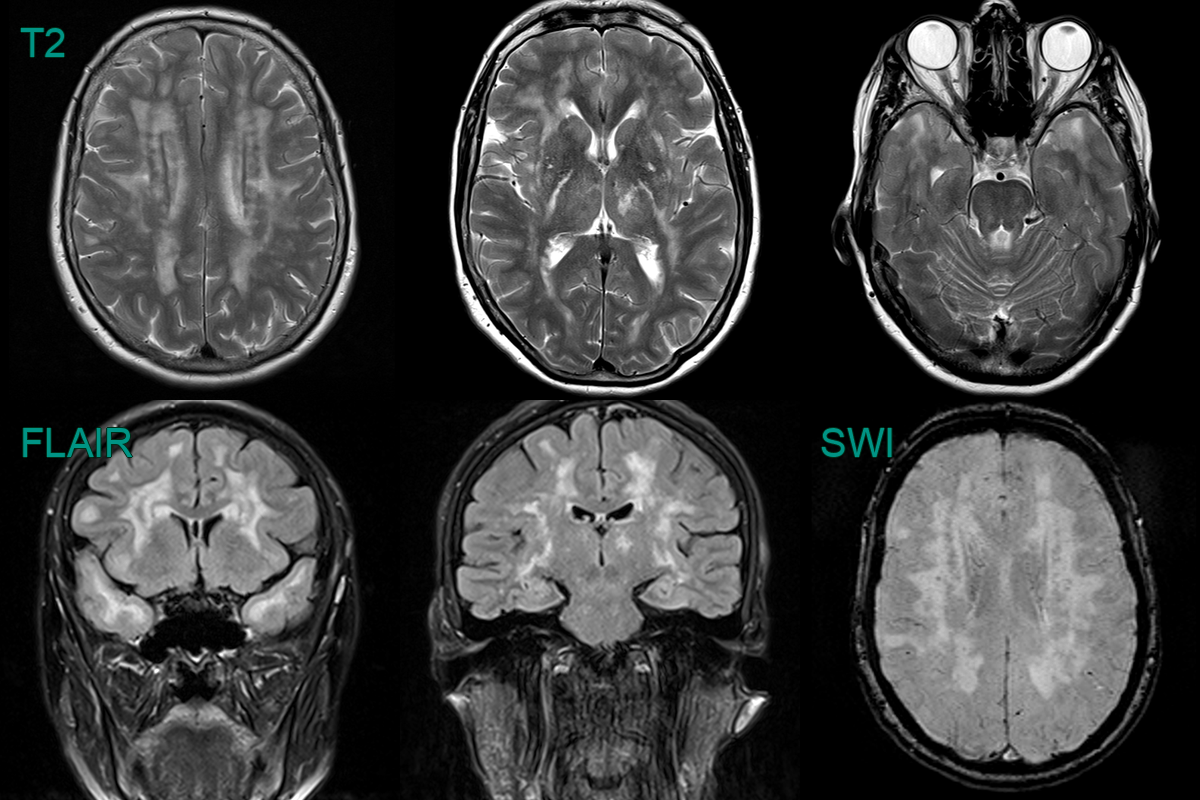
- A 55-year-old patient with no cardiovascular risk factors presented with recurrent headache.
- MRI showed a confluent leukoencephalopathy, involving the external capsules and anterior temporal lobes, and lacunar infarcts.
- There were deep and lobar microhaemorrhages.
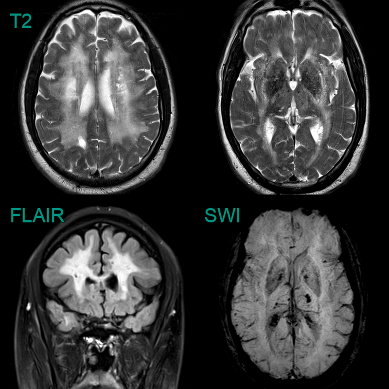
- 50-year-old patient with no cardiovascular risk factors had recurrent strokes.
- MRI showed a severe burden of small vessel disease, multiple lacunar infarcts and mixed distribution microhaemorrhages.
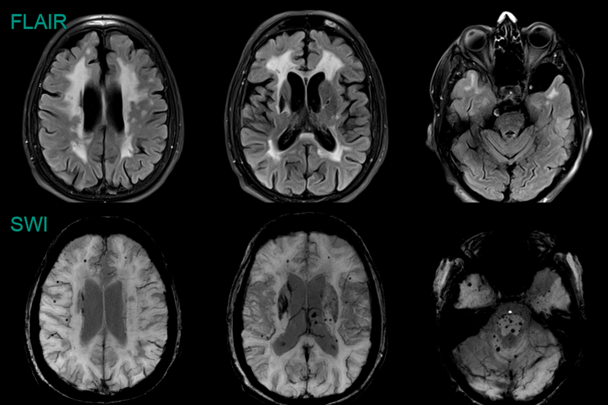
- 50-year-old patient presented with a homonymous hemianopia and mixed sensory and motor deficits.
- MRI showed an acute left thalamic infarct and a diffuse leukoencephalopathy that involved the external capsules and anterior temporal lobes.
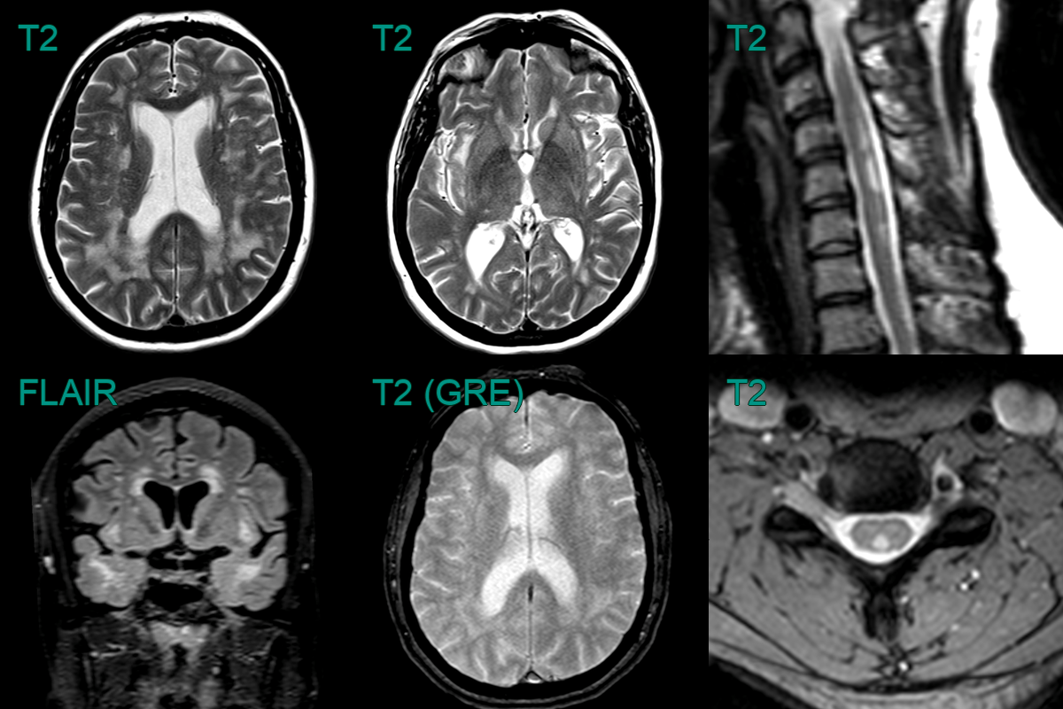
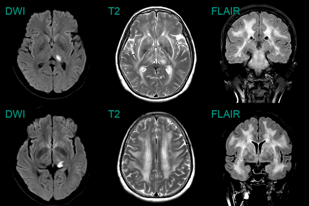
- Alongside the classical intracranial features of CADASIL, MRI showed multiple dorsal column hyperintensities.
- With no evidence of a metabolic or demyelinating cause, findings were ascribed to a CADASIL-related myelopathy.