Cerebral metastasis
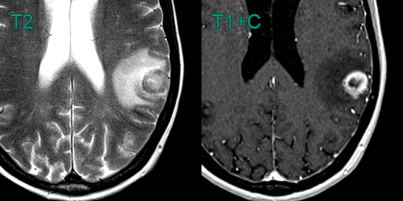
- A 60-year-old patient presented with right sided weakness.
- MRI showed a ring-enhancing lesion near grey-white matter interface with surrounding vasogenic oedema.
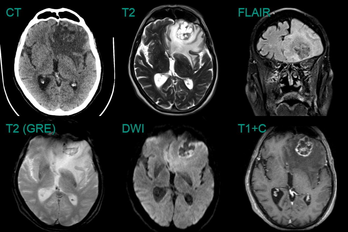
- A 80-year-old patient with a history of bladder cancer presented with headche.
- Imaging showed a peripherally enhancing lesion containing blood product in the left frontal lobe with a large volume of surrounding vasogenic oedema.
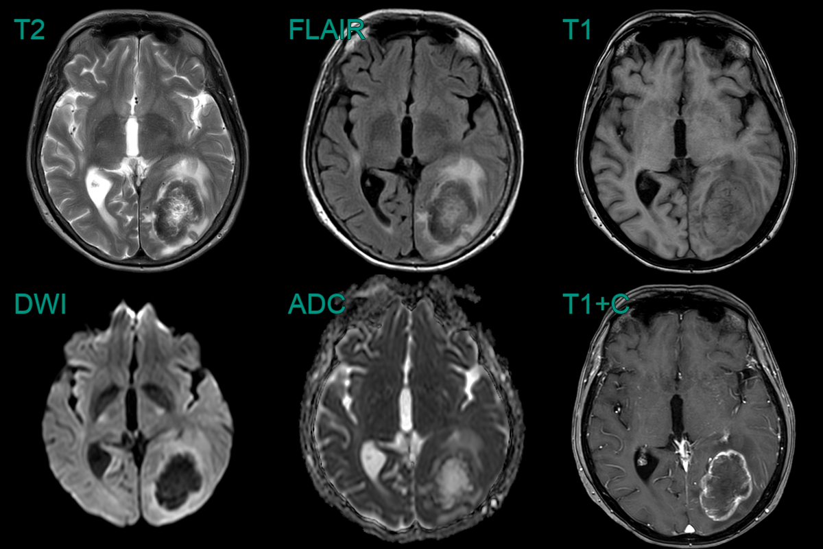
- A 60-year-old patient who was diagnosed with colonic cancer three years prior presented with a right visual field defect and headache.
- A large left occipital lobe lesion showed peripheral enhancement and was surrounded by vasogenic oedema.
- The peripheral T2-hypointensity within the lesion has been reported to be related to collagen accumulation.
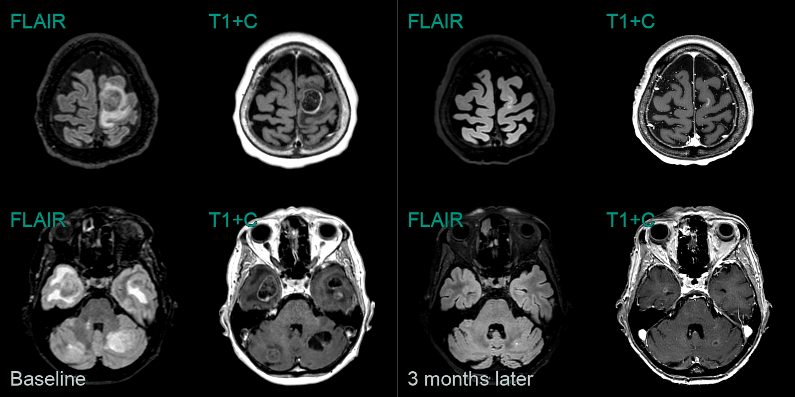
- A 70-year-old patient with small cell lung cancer presented with right leg weakness.
- MRI showed many peripherally enhancing lesions, the larges of which was in the left paracentral lobule.
- Following chemotherapy, MRI showed a marked reduction in the size of all of the lesions and the surrounding oedema.
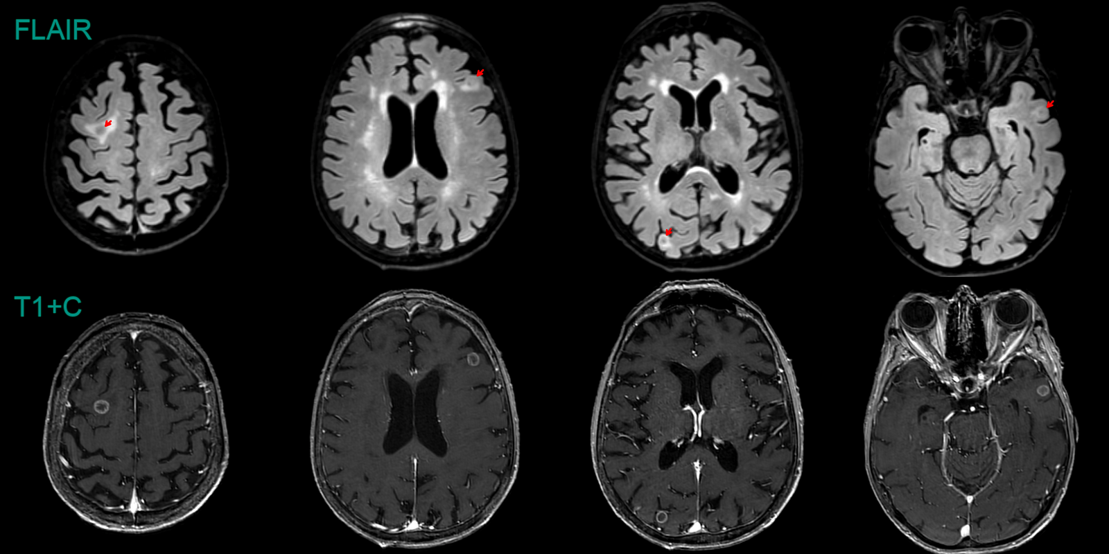
 * 70-year-old patient with a new diagnosis of non-small cell lung cancer had an MRI to 'screen' for metastasis. * MRI showed subcentrimeter ring-enhancing lesions with variable amounts of surrounding vasogenic oedema.