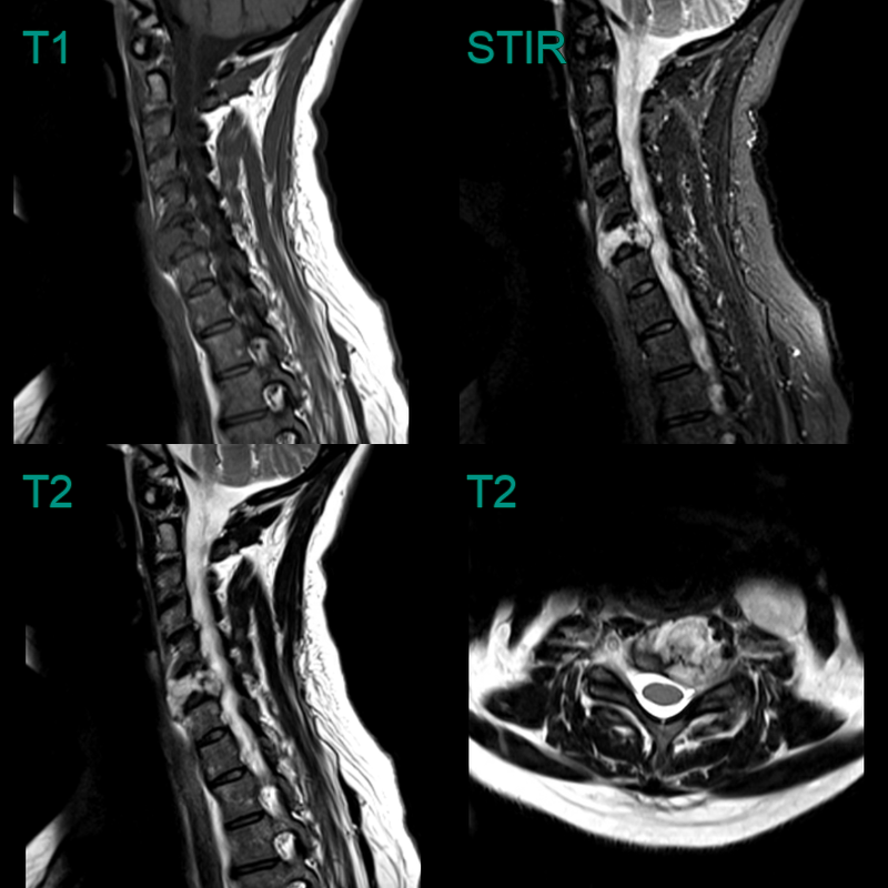
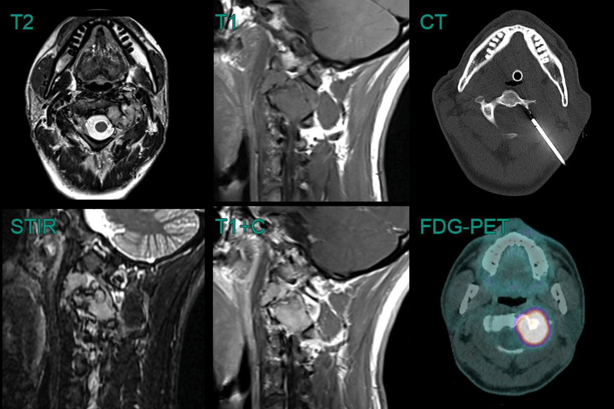
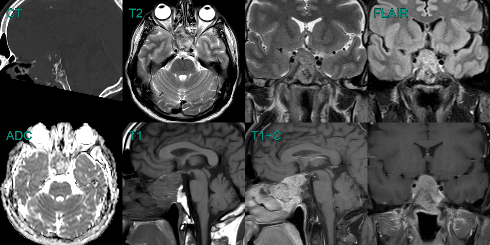
Chordoma
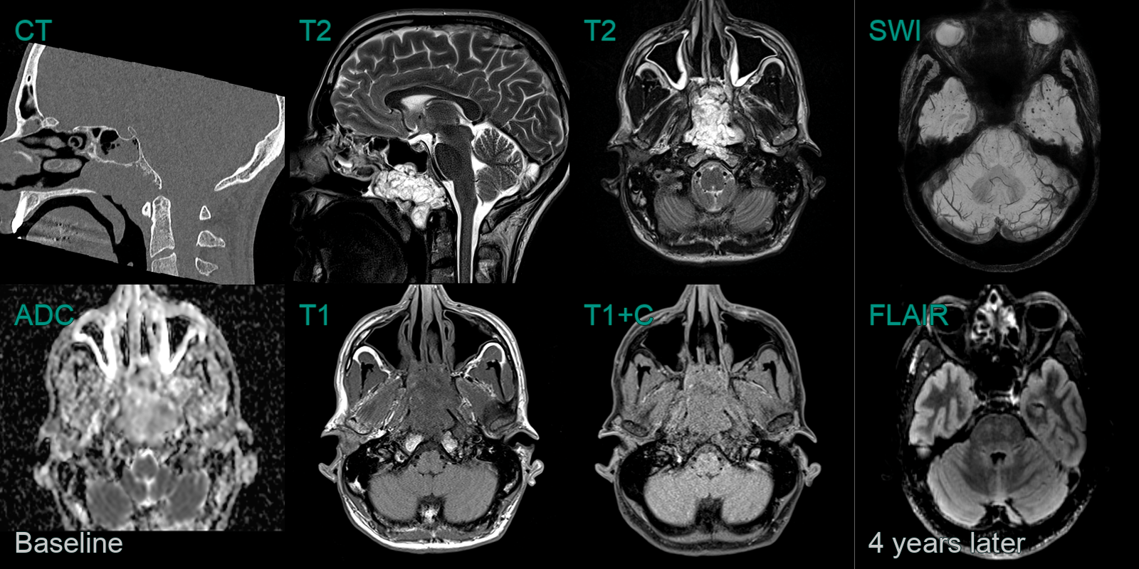
- A 50-year-old patient presented with nasal obstruction.
- MRI showed a lobulated mild enhancing lesion in the nasopharynx with erosion of the inferior cortex of the clivus.
- 4 years later, a follow-up MRI showed no recurrence but many microhaemorrhages in the anterior temporal lobes and brainstem, which were likely to be related to radiotherapy.
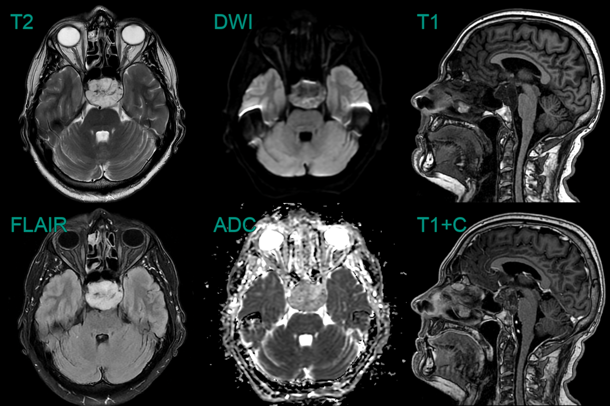
- A 50-year-old patient presented with a visual field defect picked up during a routine eye test.
- CT showed a large destructive lesion centred on the anterior clivus and pituitary fossa.
- MRI showed an avidely enhancing clivus lesion that was compressing the right cisternal optic nerve.
- With the differential including a pituitary macroadenoma, a chordoma was confirmed following a transphenoidal biopsy.
