Dermoid cyst
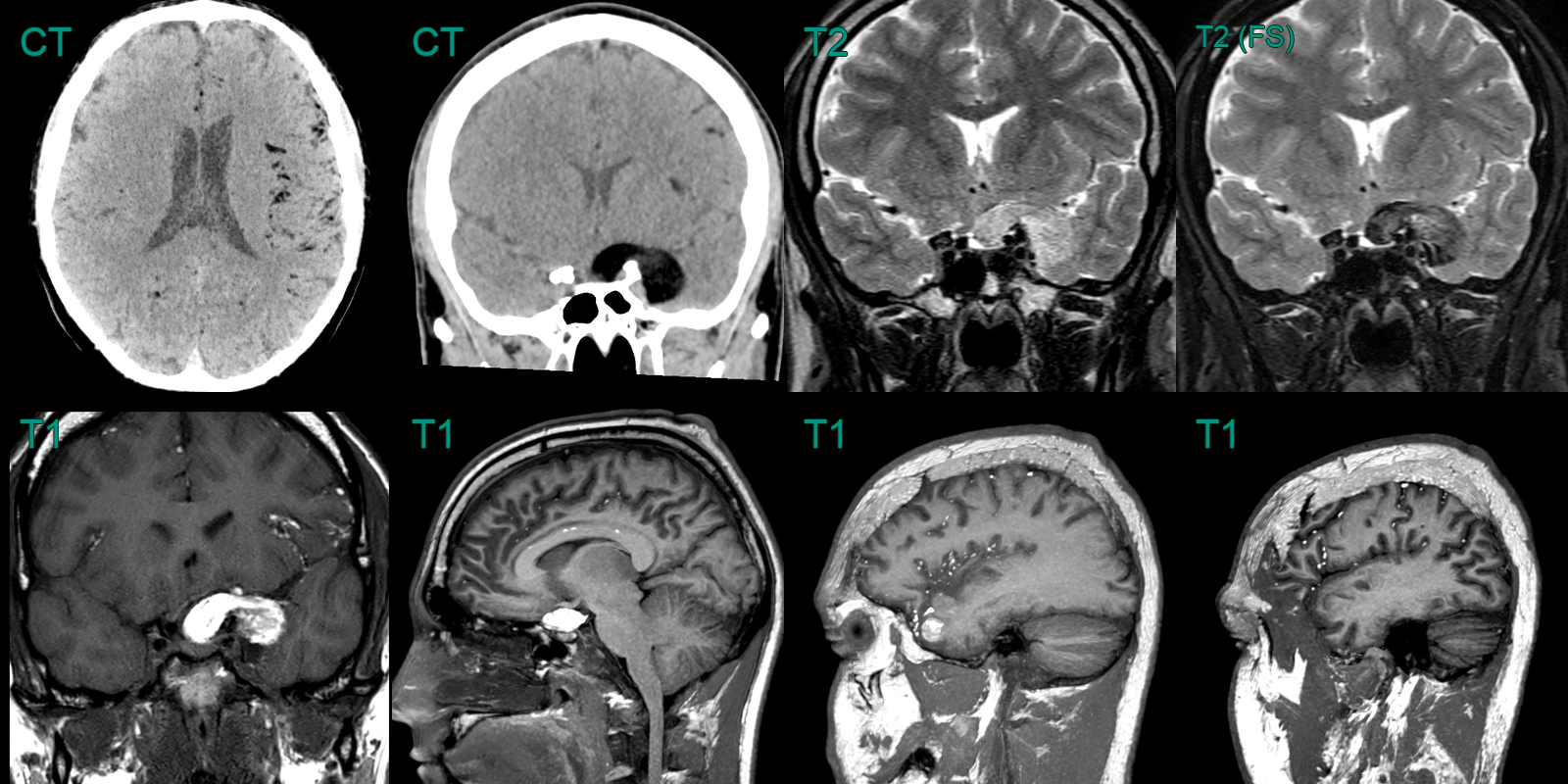
- A 30-year-old patient presented with an acute onset headache.
- MRI showed a lesion in the right cavernous sinus that was T1-hyperintense that suppressed on the fat-suppressed FLAIR imaging, consistent with fat content.
- There were further locules of fat signal over the cerebral hemispheres consistent with dermoid cyst rupture.

