Dural venous sinus thrombosis
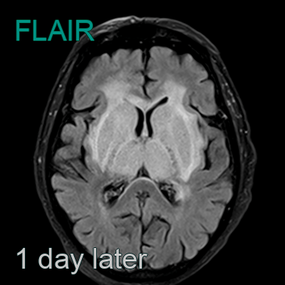
- 65-year-old patient on treatment for metastatic lung cancer.
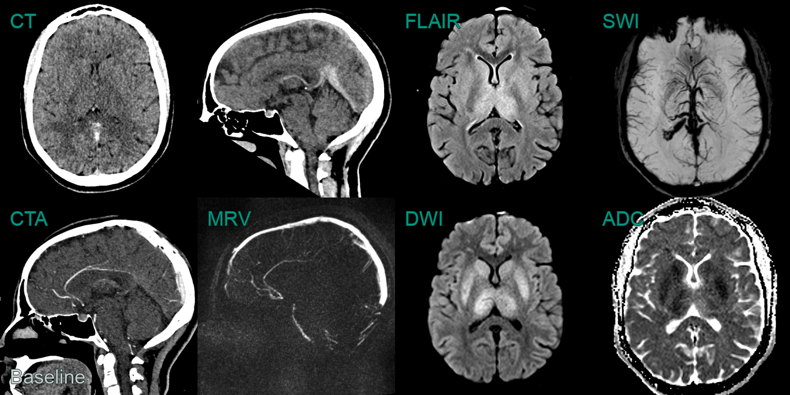
- CT showed hyperdensity in the straight sinus, vein of Galen and internal cerebral veins.
- These vessels did not enhance on the CTV.
- MRI the next day showed hyperintensity and swelling of the deep grey structures and the capsular white matter.
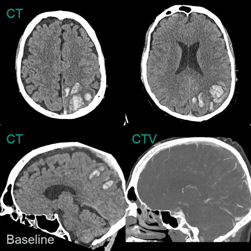
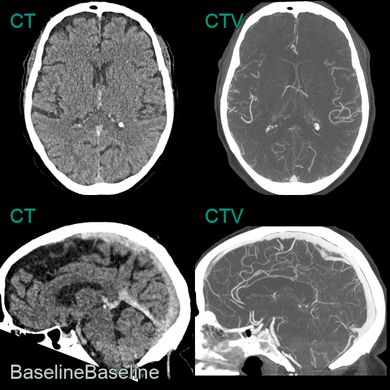
- 50-year-old patient presented with headache and dysphasia.
- CT showed a hyperdense superior sagittal sinus and a lobulated, fractionated, haematoma in the left parietal and occipital lobe.
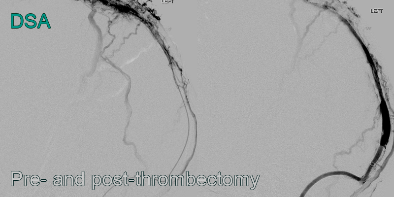
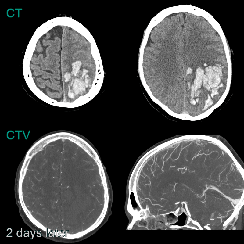
- Despite a successful venous thrombectomy, thrombus reaccumulated in the superior sagittal sinus and the haematoma enlarged.

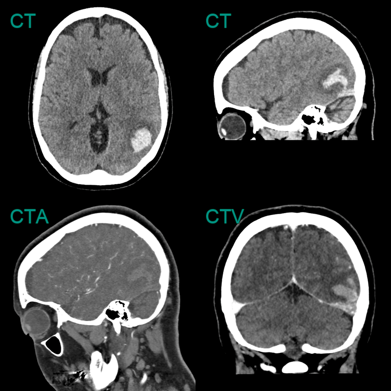
- 30-year-old patient presented with headache, confusion and expressive dysphasia.
- CT showed subtle hypodensity in the deep grey nuclei and hyperdensity within the straight sinus, vein of Galen, and internal cerebral veins. These structures were occluded on both the CTA and phase-contrast MRV.
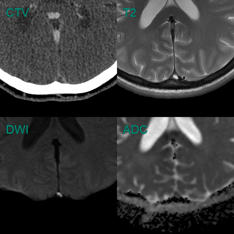
- MRI showed hyperintensity and diffusion restriction in the deep grey nuclei. SWI showed congenstion of the deep venous system.
- 2 weeks later, a repeat MRI showed only very minimal hyperintensity in the thalami (where some microhaemorrhages had developed) indicating that the diffusion restriction was largely reversible although a few microhaemorrhages developed in the thalami.
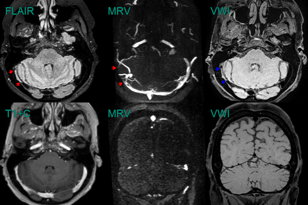
- A 35-year-old patient presented 3 months after the onset of a headache.
- FLAIR showed a hyperintense segment in the right transverse sinus with absent flow on the MRV (red arrows).
- While thrombus was strongly suspected, this was confirmed with black blood imaging (blue arrows), removing the possibility of the findings on FLAIR and MRV were artefactual.
- Notice that on the post-gadolinium images, the filling defect is not apparent due to thrombus enhancement.