Enlarged perivascular spaces
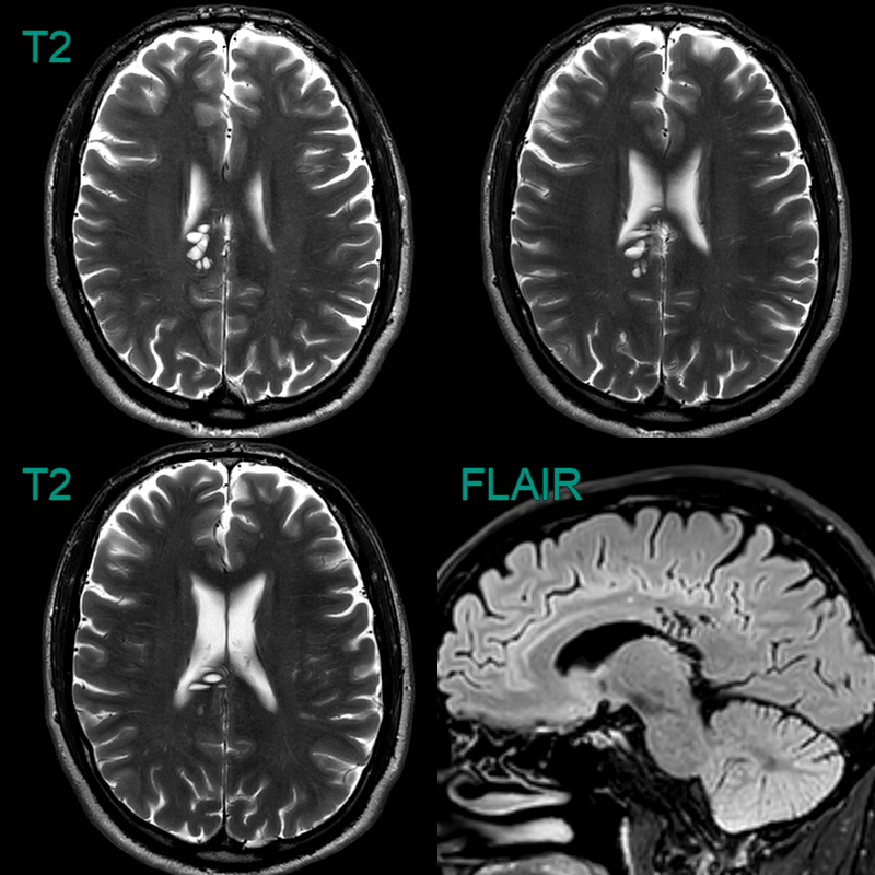
- A cluster of T2-hyperintense lesions centred on the right cingulum fully suppressed on FLAIR and had no surrounding parenchymal signal change.
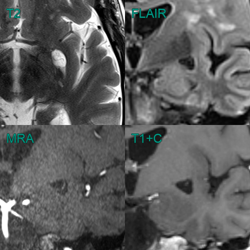
- There is an enlarged perivascular space (T2-hyperintense and fully suppressing on FLAIR) in a typical location; in the subganglionic region.
- Both T2-weighted, time-of-flight angiography and post-gadolinium T1-weighted imaging showed the vessel traversing the perivascular space.
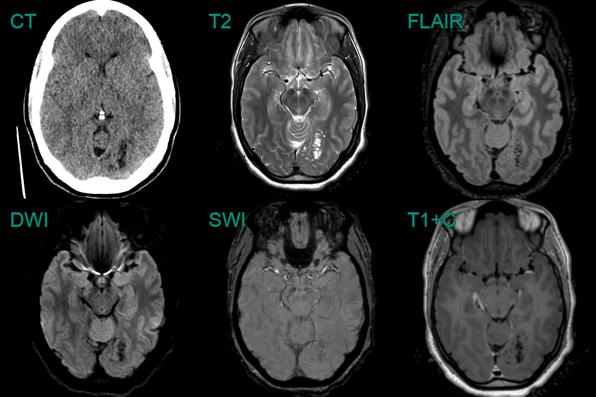
- Incidental finding of a clustered subcortical CSF signal without mass effect was consistent with enlarged perivascular spaces.
- With no FLAIR hyperintensity, an MVNT was not likely.
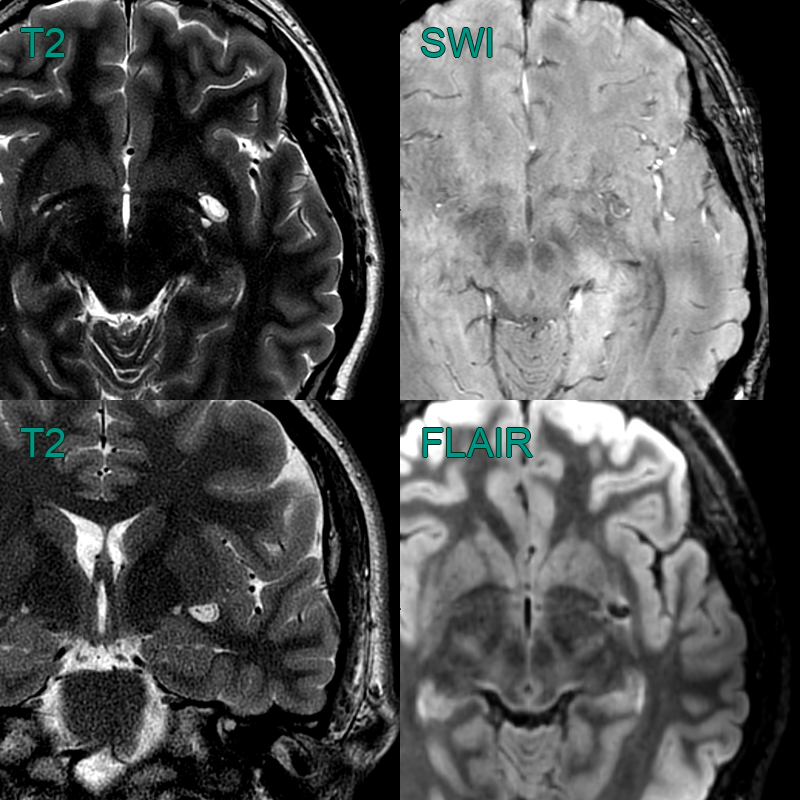
- An incidental enlarged perivascular space in the left subganglionic region.
- The transiting artery is seens as a flow void on T2-weighted imaging.