Epidermoid cyst

panels-1

panels-1

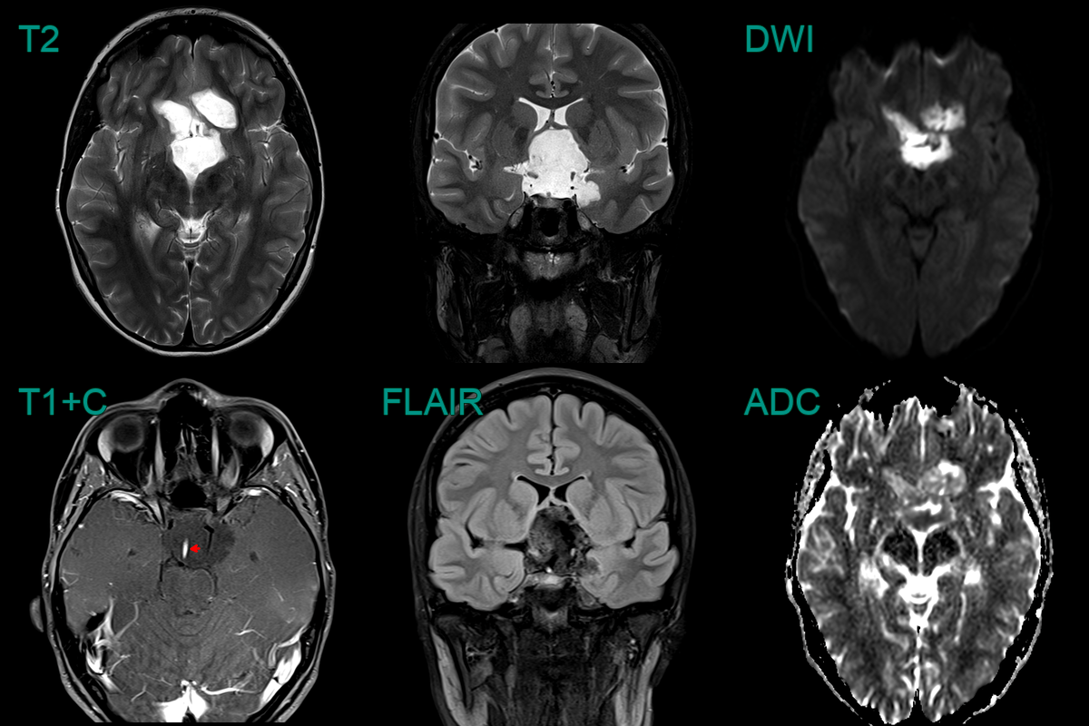
  • 25-year-old patient presented with headache.
  • MRI showed a diffusion-restricting non-enhancing suprasellar lesion that encased arteries and the infundibular stalk.

panels-1

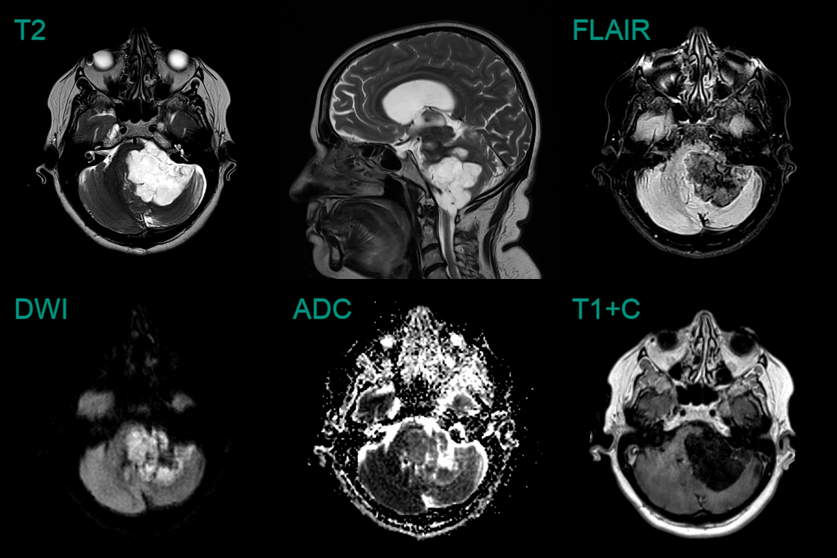
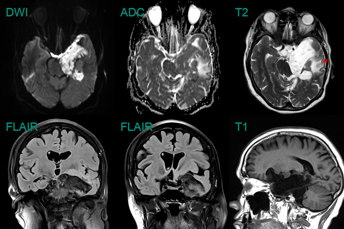
  • 60-year-old patient present with ataxia and poor left-sided hearing.
  • MRI showed a T2-hyperintense non-enhancing lobulated lesion with low ADC values in the left side of the posterior fossa, encasing the 7th and 8th nerve complexes.
  • There was significant mass effect on the cerebellum (presumably relevant to the ataxia) but there was no oedema, indicating that this lesion has grown slowly.
panels-1

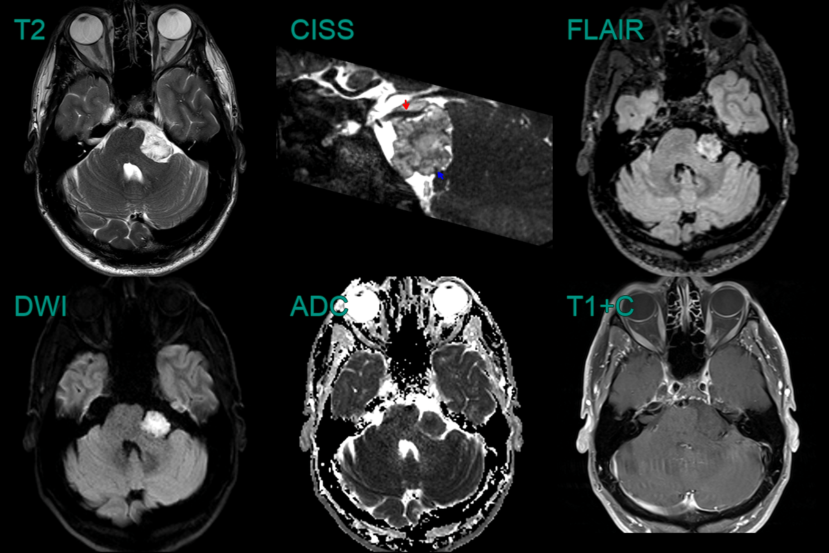
  • 40-year-old patient presented with facial weakness and paraesthesia.
  • MRI showed a 2.2 cm lesion in the left cerebellopontine angle cistern that caused diffusion restriction.
  • The lesion displaced the trigeminal nerve (red) and facial nerve (blue).