Fibrous dysplasia
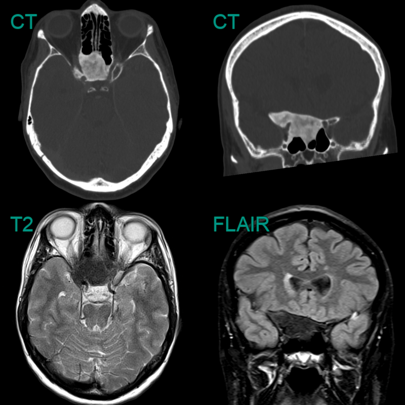
- A 50-year-old patient complained of a hard lump behind there left ear.
- CT showed a heterogeneously expanded left occipital bone.
- MRI showed the lesion to have mixed signal intensity on diffusion-weighted and T2-weighted imaging and patchy enhancement.
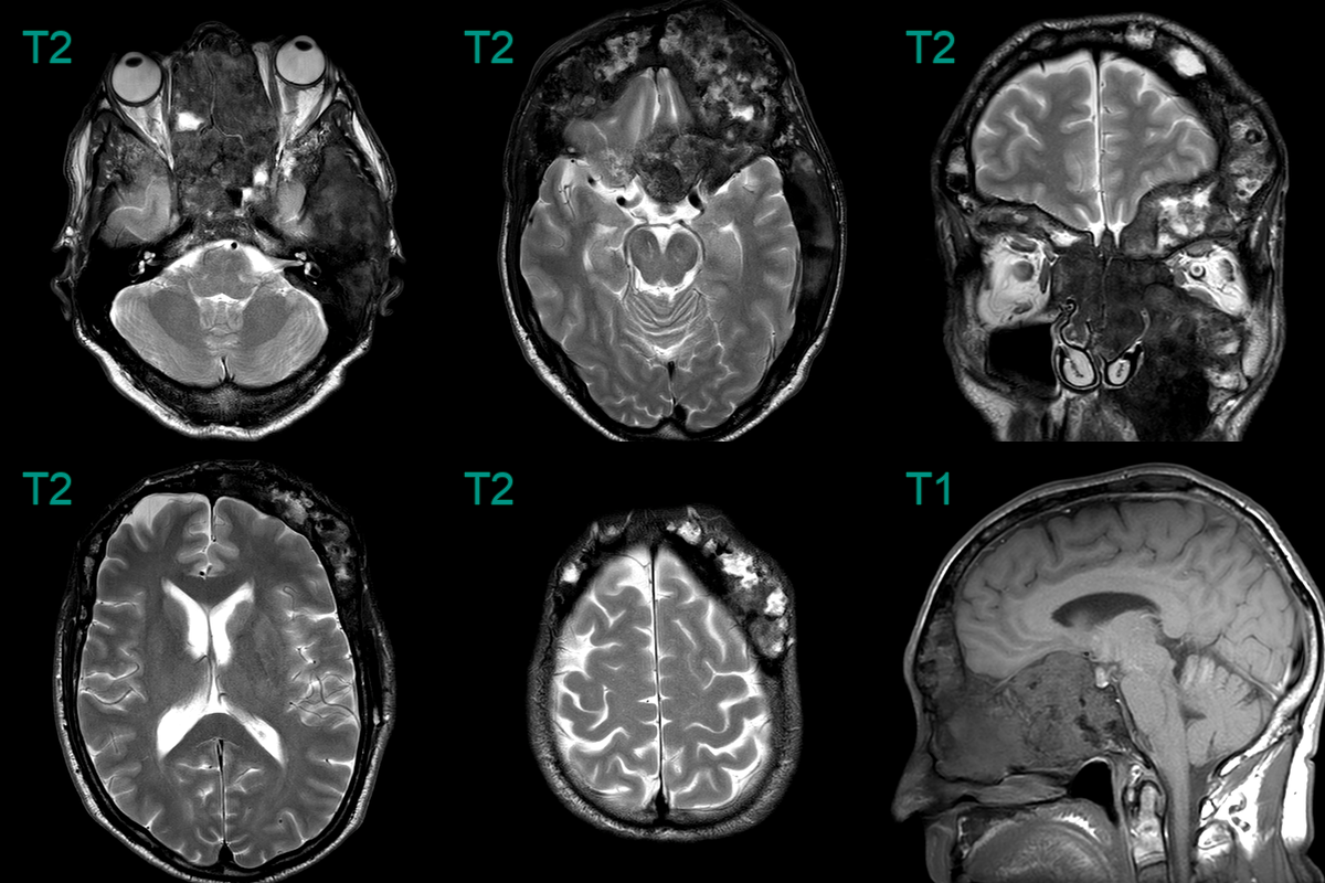
- Fibrous dysplasia involving multiple bone of the skull in the context of McCune-Albright syndrome.
- The expansile lesion in the sphenoid bone has a ground-glass texture on CT.
- On MRI, the lesion was hypointense on all sequences.
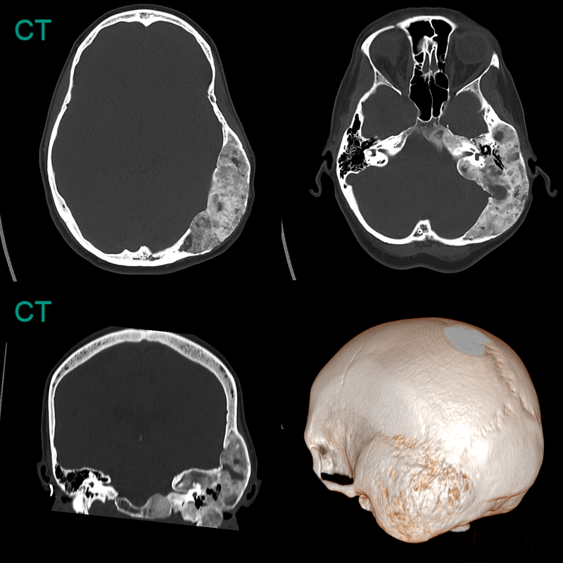
- 45-year-old patient presented with a longstanding skull deformity.
- CT showed an expansile lesion centered on the left temporal bone with the ground glass texture that is classic for fibrous dysplasia.
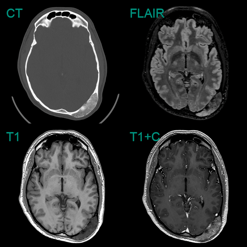
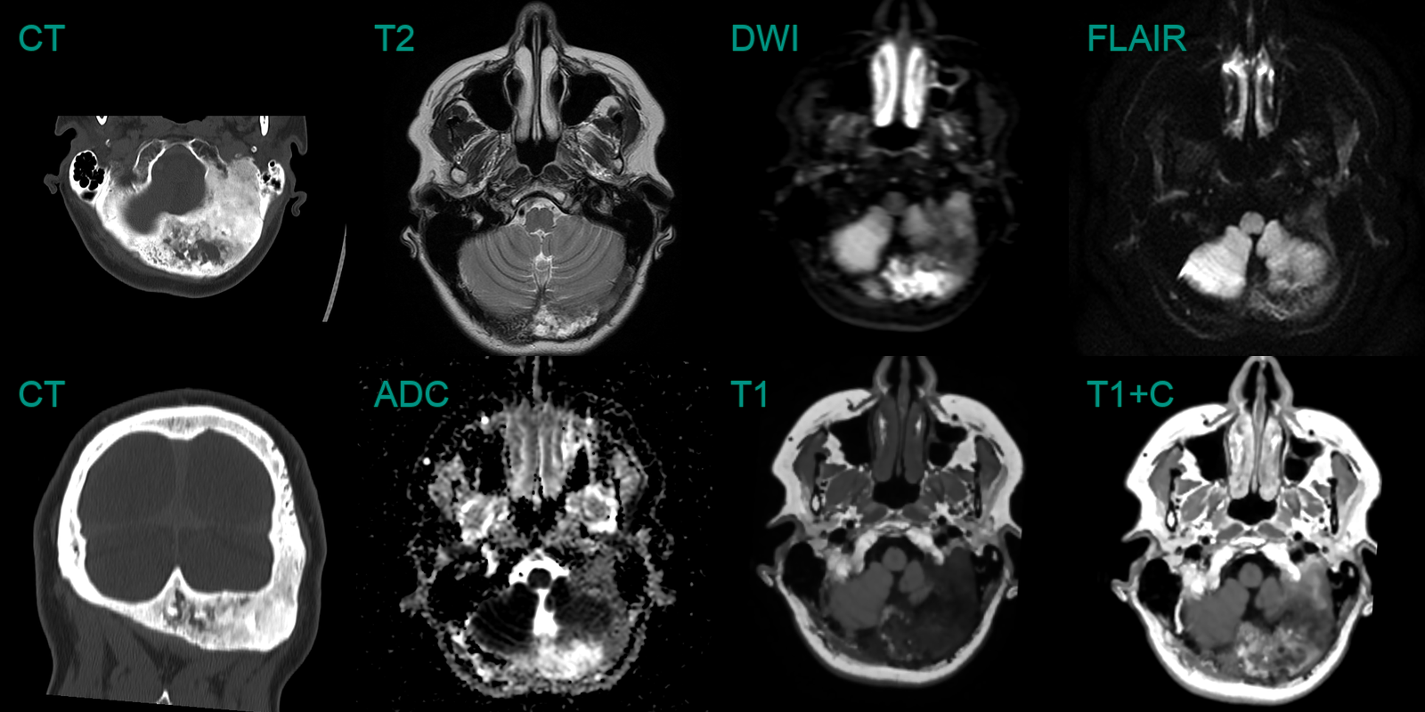
- A 50-year-old patient presented with a painless lump over the back of the head.
- Imaging showed a hazy well-demarcated low-density lesion with heterogenous enhancement consistent with fibrous dysplasia.