Glomus tympanicum
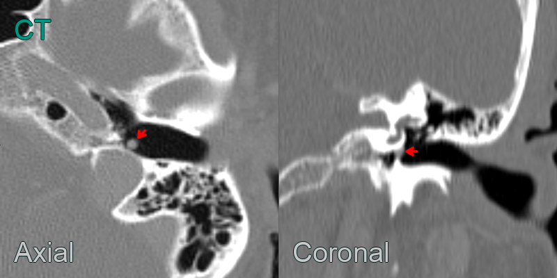
- 60-year-old patient presented with left sided pulsatile tinnitus.
- Cone beam CT showed a 3 mm nodule interposed between the hypotympanic jugular bulb and the caudal aspect of pars tensa.
- A gloums tumour was confirmed following resection.
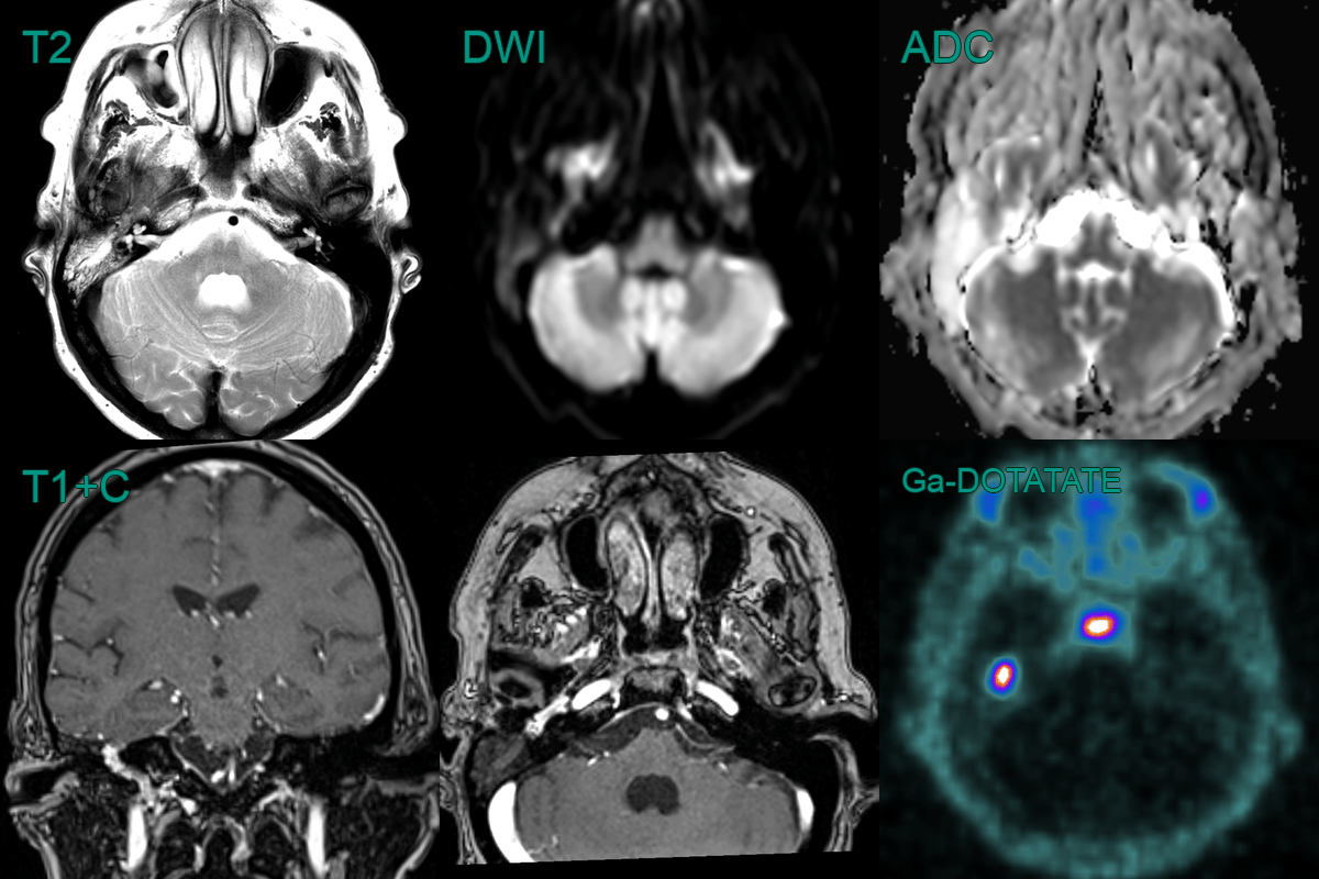
- A 70-year-old patient presented with right sided pulsatile tinnitus.
- Otoscopy revealed a dark lesion associated with the tympanic lesion.
- MRI showed an avidely enhancing lesion deep to the tympanic membrane extending into the hypotympanum.
- Ga-DOTATATE PET showed high avidity in the lesion, consistent with a glomus tumour.