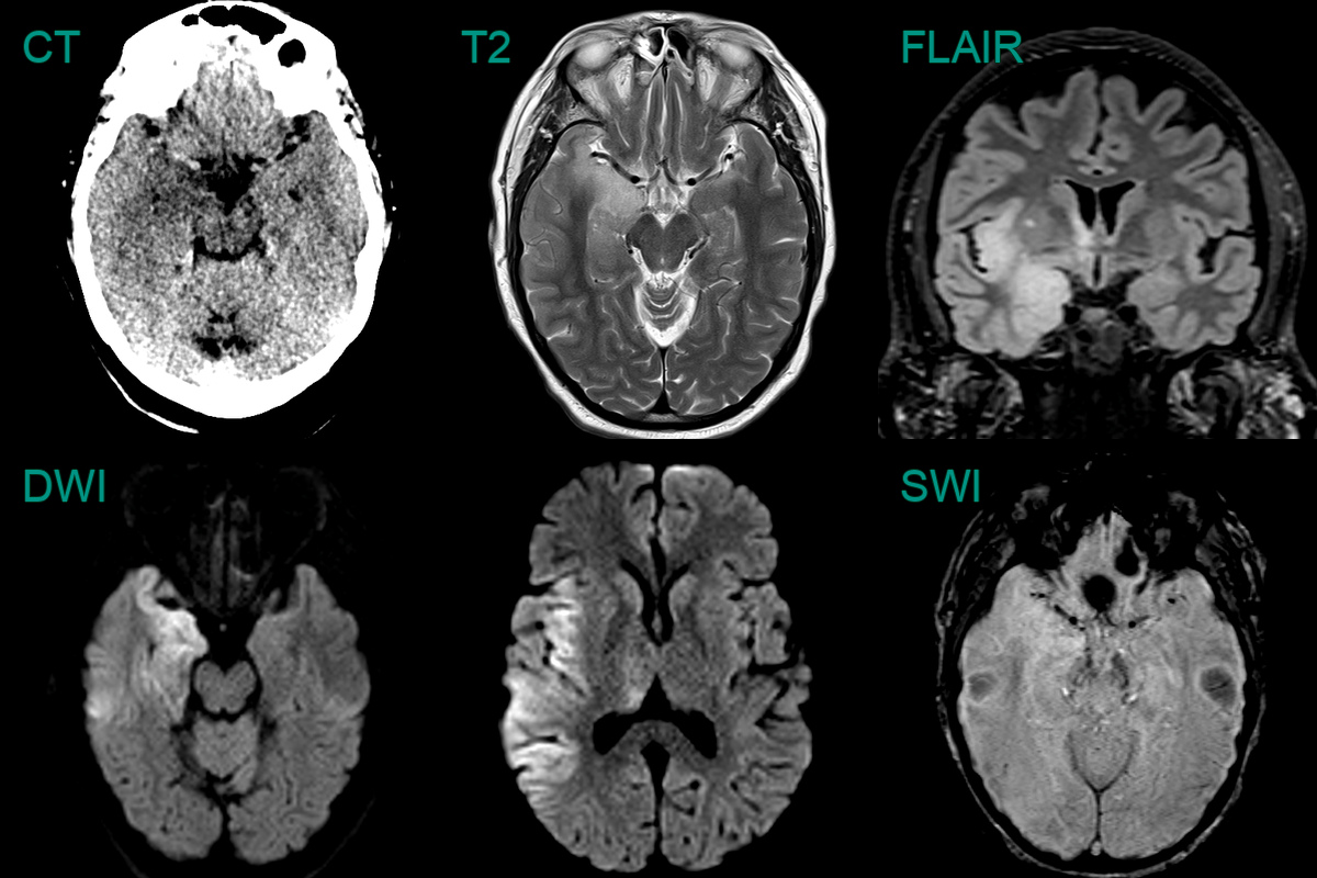
50-year-old patient presented with headache and confusion.
CT showed swelling and low attenuation in the anterior and mesial left temporal lobe and in the right hippocampus.
The parenchymal low attenuation gave the impression of a hyperdense vessel (Mach effect) but infarction was not likely as both MCA nad PCA territories were involved.
There was high T2, FLAIR and DWI signal (without clear diffusion restriction) but no enhancement.
40-year-old patient presented with 2 day history of confusion and reduced GCS, headache and fever.
MRI showed hyperintensity and swelling of the right mesial temporal lobe and diffusion restriction extending up to the right parietal lobe.
70-year-old patient presented with dysphasia and right-sided weakness and seizures. HSV was identified in CSF.
MRI showed diffusion restriction in the cortex of left cerebral hemisphere as well as the right insula. There was swelling and subtle cortical enhancement.
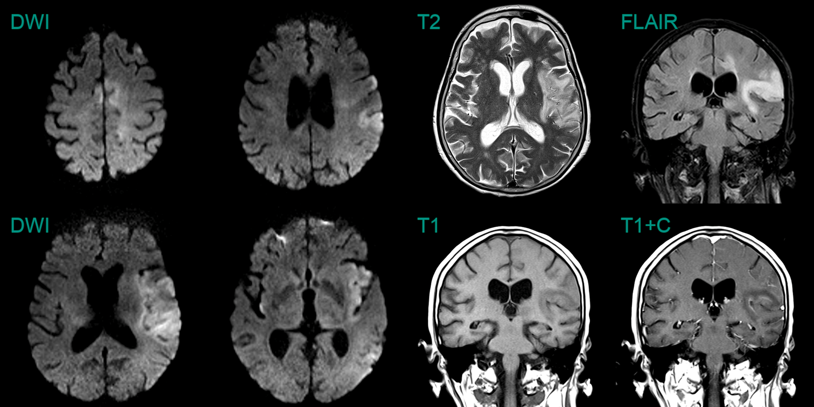
60-year-old patient presented with expressive dysphasia.
MRI showed diffuse patchy cortical, white matter and ganglionic hyperintensity.
On follow-up, hyperintensity involving most of the left temporal lobe resolved while marked hyperintensity developed in the right temporal lobe.
Despite being repeatly negative on CSF, brain biopsy reavealed an HSV encephalitis.
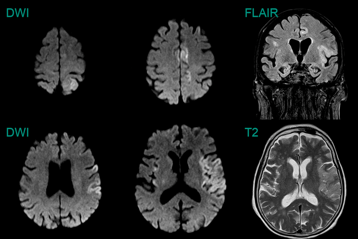
75-year-old patient presented with dysphasia and right sided weakness.
MRI showed cortical DWI hyperintensity in the left frontal, parietal, temporal lobes and insular cortex bilaterally.
Initially concerned about an acute infarct, the widespread and exclusively cortical involvement and a normal CTA, made HSV encephalitis more likely (which was confirmed on CSF analysis).
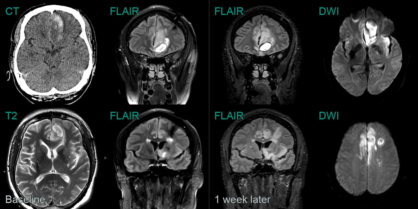
A 45-year-old patient presented with right sided weakness and a fever.
CT showed a haematoma in the left paramedian frontal lobe.
MRI showed oedema around the haematoma as well as subtle high FLAIR signal in the cortex of the left frontal lobe and the right cingulate.
On follow-up imaging, the FLAIR hyperintensity (and diffusion restriction) had extended through the limbic system.