Intracranial lipoma
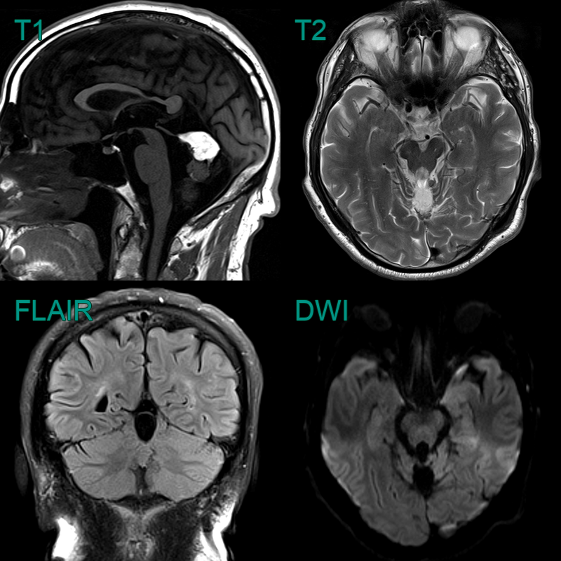
- T1-hyperintense lesion in the posterior fossa that suppressed on fat-suppressed FLAIR imaging representing a a fat-filled lesion. With no non-fat components (and no change in 10 years), the lesion is most consistent with a lipoma.
- The lipoma was associated with a small volume (or partially absent) vermis; this could be due to hypoplasia rather than atrophy.
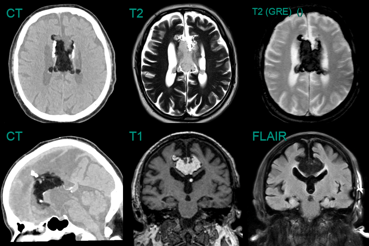
- An incidental fat-containing lesion contained calcification and was associated with a posteriorly deficient corpus callosum.
- The lesion contained fat based on T1 hyperintensity and suppression of signal of fat suppressed sequences (FLAIR and GRE T2).