Intraosseous haemangioma
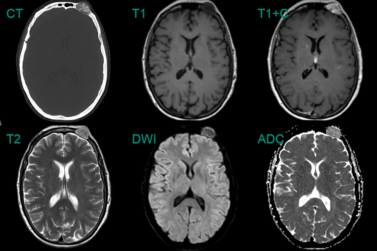
- A 40-year-old patient presented with an enlarged forehead lump.
- Imaging showed a partially calcified avidly enhancing intraosseous lesion in the left frontal bone.
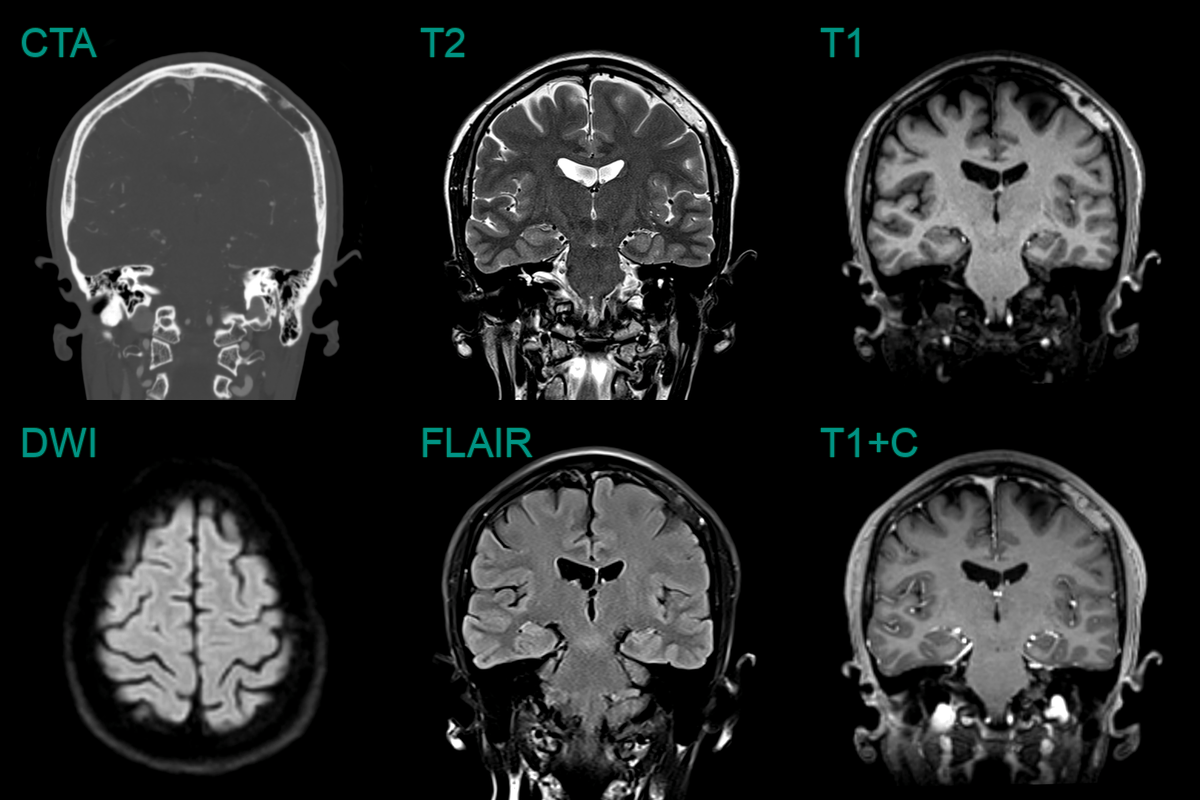
- An incidental lesion in the intradiploic space of the parietal bone contained fat and did not enhance (allowing for prominent nearby veins).
- The bone appearance was not typical, but a radiological diagnosis of a fatty haemangioma was given. A lipomatous lesion is also possible.
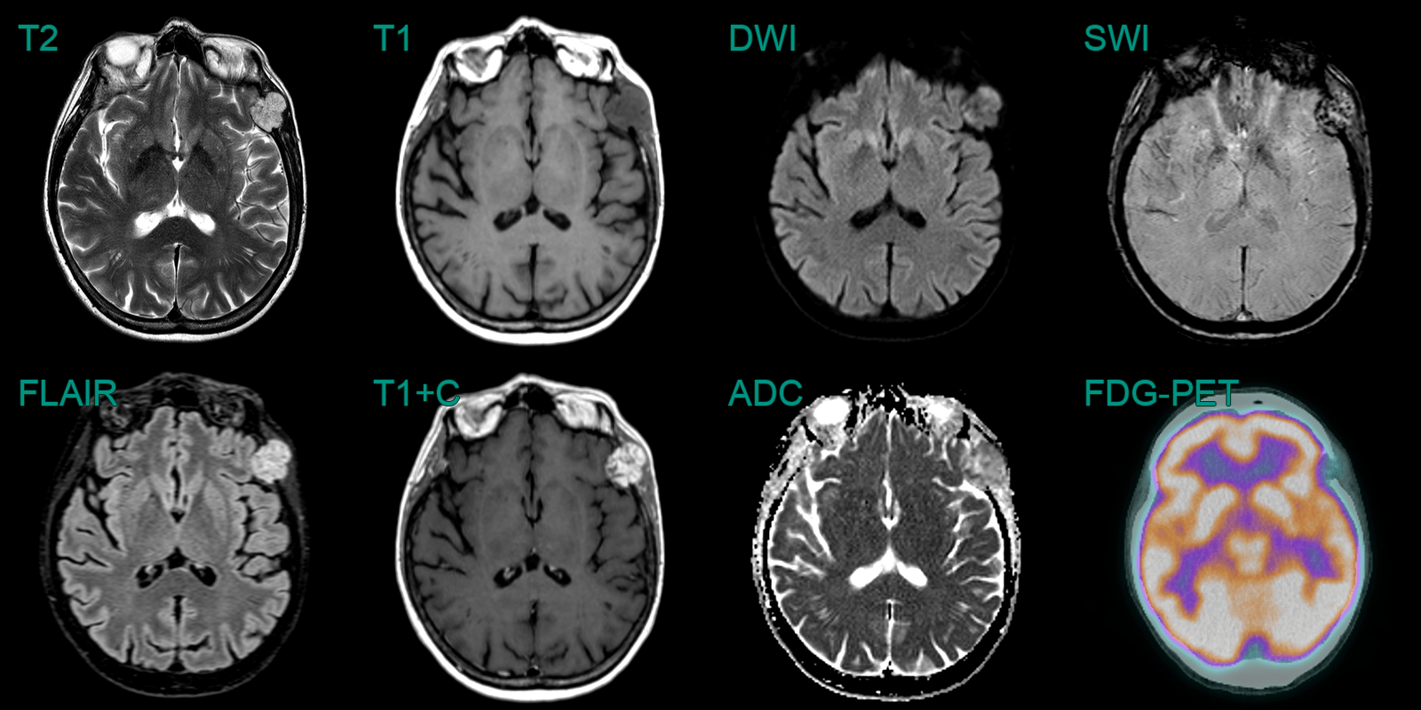
- A 70-year-old patient presented with a lump near the left zygoma.
- MRI showed an avidly enhancing lesion replacing part of the frontal bone without significiant avidity on FDG PET.
- Histopathology following resection (due to slow enlargement) confirmed a haemangioma.