Mitochondrial Encephalopathy with Lactic Acidosis and Stroke-like Episodes (MELAS)


- 40-year-old patient presented with acute onset of aphasia.
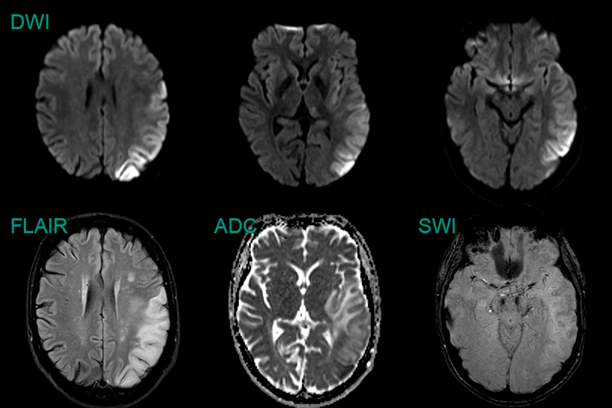
- Initial MRI showed cortical and subcortical diffusion-weighted abnormality in the left anterior temporal lobe.
- On follow-up imaging, this region of signal abnormality and swelling significantly increased. There was both restricted and facilitiated diffusion involving both cortex and subcortical white matter.
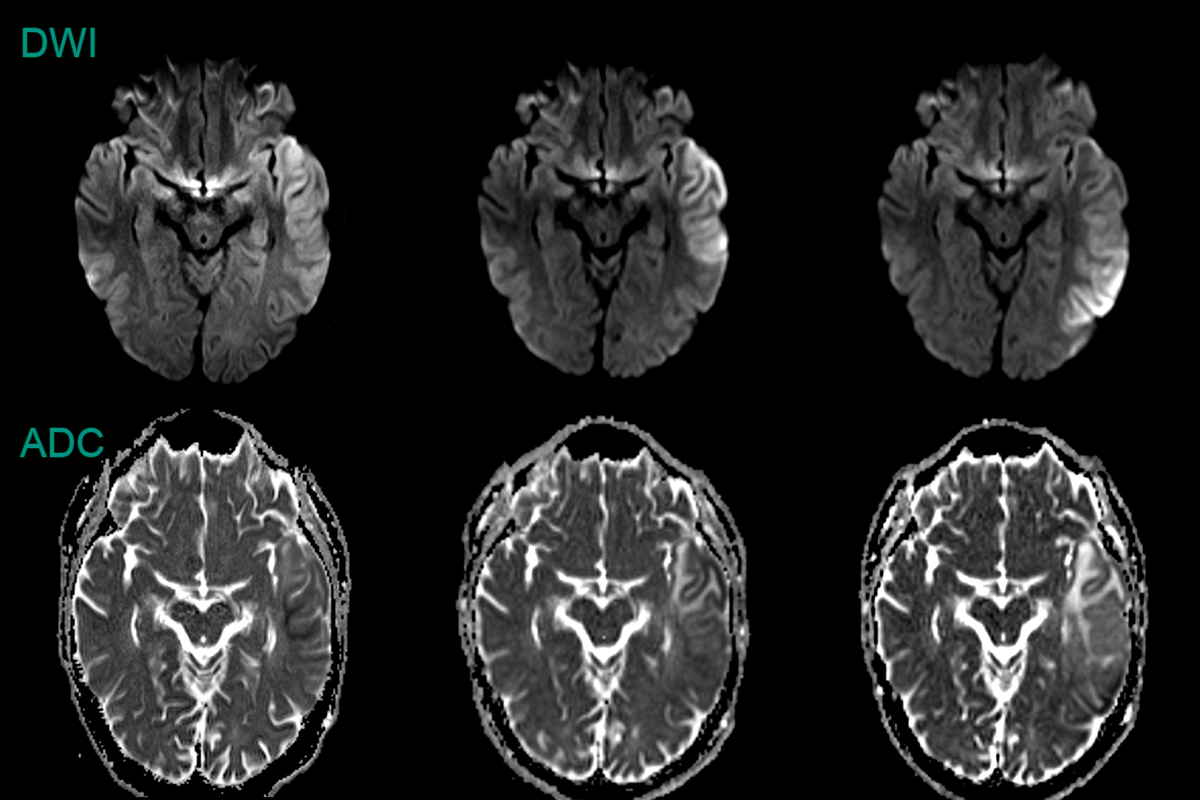
- A 30-year-old patient presented with a 1 week history of word finding difficulty followed by a seizure.
- MRI showed cortical diffusion restriction variably affecting the cortex and subcortical white matter of the left cerebral hemisphere.
- The region of abnormality increased over the 3 weeks.