Meningioma
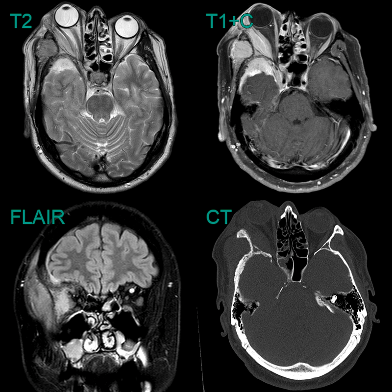
- Patient presented initially presented with headache and proptosis.
- CT showed mixed hyperostosis and luceny of the right sphenoid bone.
- MRI showed enhancing soft tissue in the middle cranial fossa, temporal fossa, and orbit (causing proptosis).
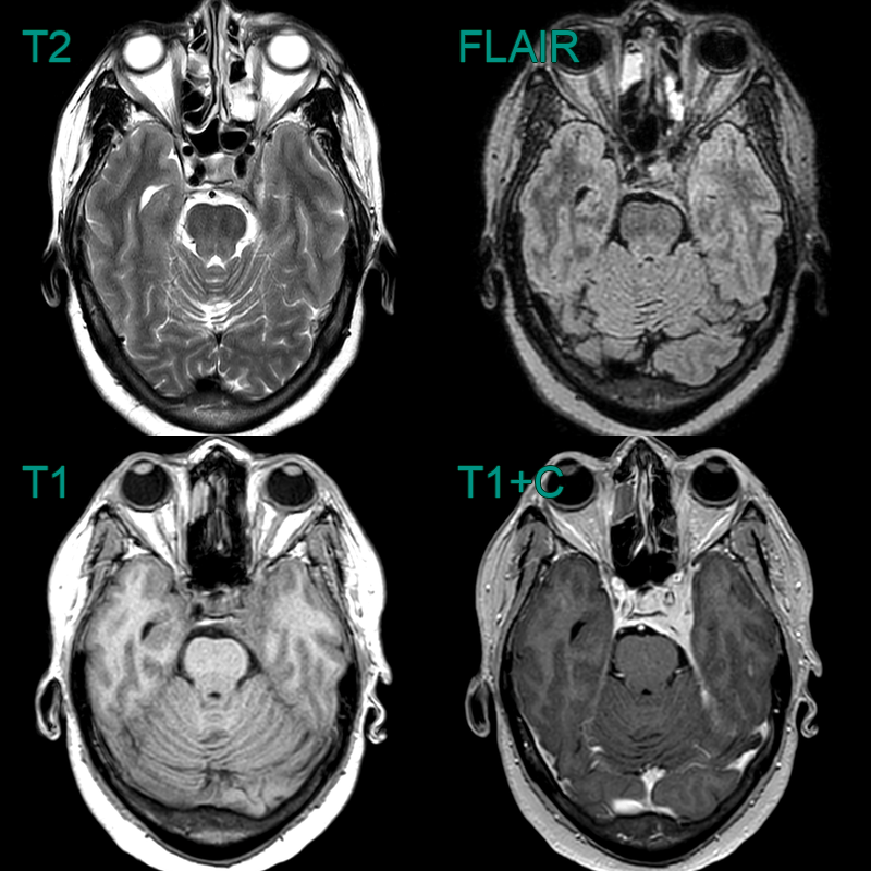
- 60-year-old patient had an MRI following trauma.
- A incidental enhancing dural lesion lesion involving the cavernous sinus (and Meckel's cave) was consistent with a meningioma.

- 50-year-old patient presented with headache.
- MRI showed an avidly enhancing lesion containing trace amounts of calcium arising from the anterior skull base.
- The ipsilateral frontal sinus was asymetrically enlarged - representing pneumosinus dilatans.
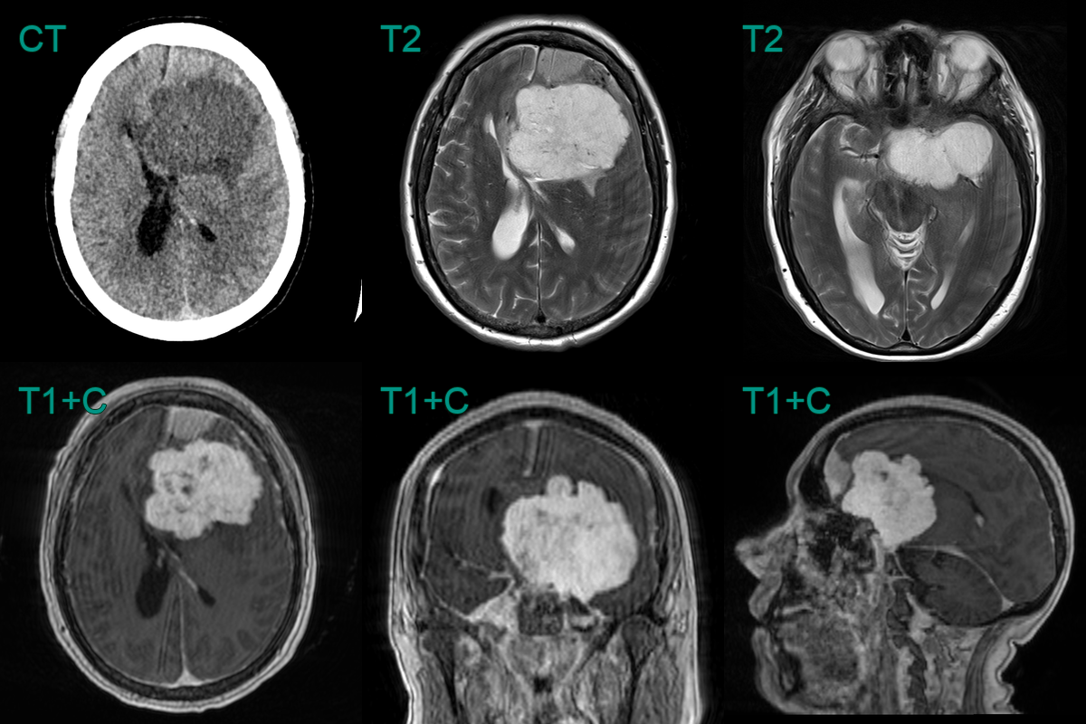
- 55-year-old patient presented with a progresively worsening headache.
- MRI showed a large avidly enahncing lesion occluding the superior sagittal sinus and eroding into the skull.
- Final histopathology revealed an atypical meningioma that was treated with radiotherapy following resection.
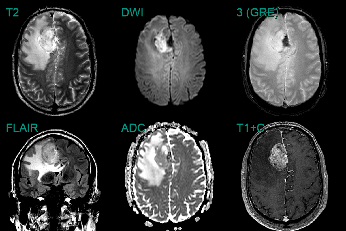
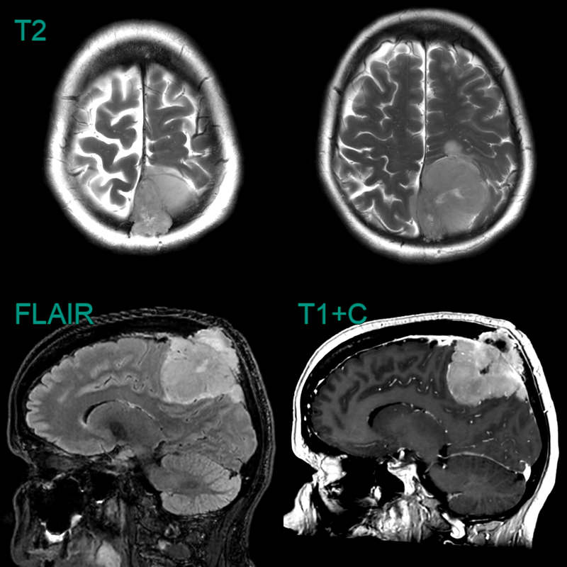
- A 50-year-old patient presented with a frontal headache.
- MRI showed a large enhancing right paramedian mass lesion associated with extensive vasogenic oedema.
- T2-hypointensity in the medial part of the lesion was caused by calcification.