Multiple Sclerosis (MS)
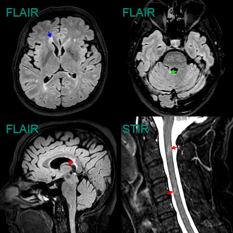 * 45-year-old patient with relapsing remitting multiple sclerosis showing lesions in the juxtacortical, periventricular, posterior fossa white matter, the corpus callosum and the cervical cord. * As there are lesions in more than one region set out in the MacDonald criteria, the imaging fulfils the criteria for dissemination in space. Dissemination in time would require either an enhancing lesion or a new lesion on a follow-up scan.
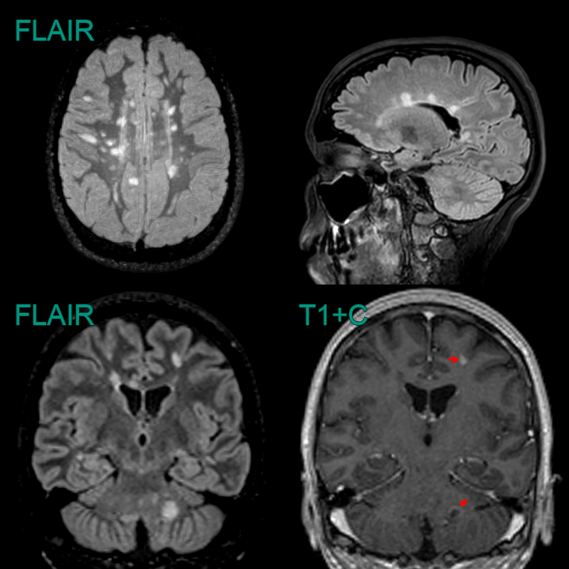
- 30-year-old patient with RR MS developed two new lesions.
- A left frontal juxtracortical lesion solidly enhanced.
- A left cerebellar lesion showed an incomplete rim of enhancement.
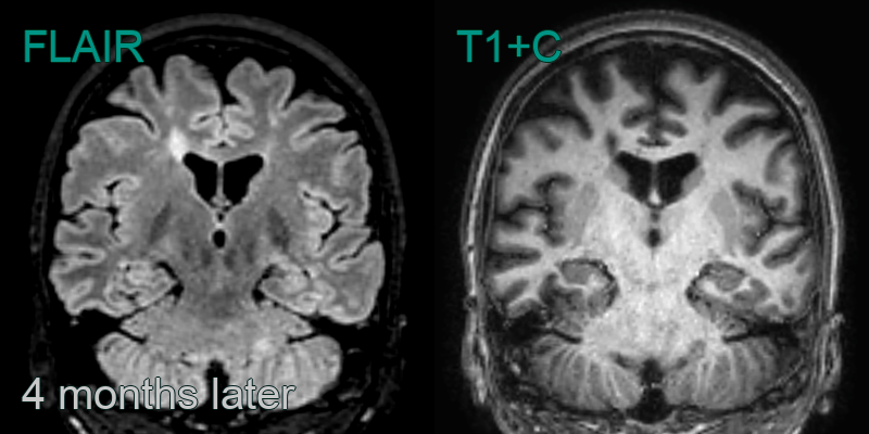
- The lesions regressed after 4 months.
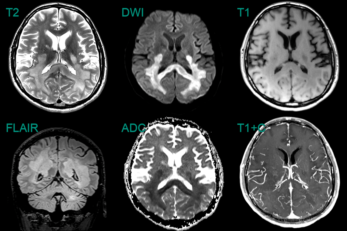
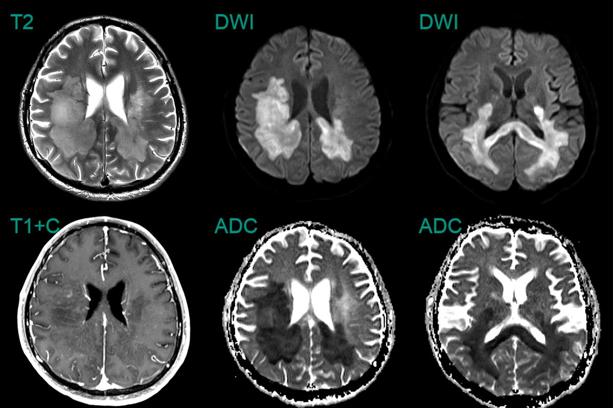
- A 30-year-old male with a known diagnosis of RRMS presented with a deterioration in motor function.
- MRI showed a large conflent diffusion restricting lesion within the posterior cerebral white matter and corpus callosum.
- The minimal contrast enhancement and only mildly elevated CBV made a lymphoma less likely.
- Biopsy confirmed tumefactive demyelination.