Optic nerve sheath meningioma
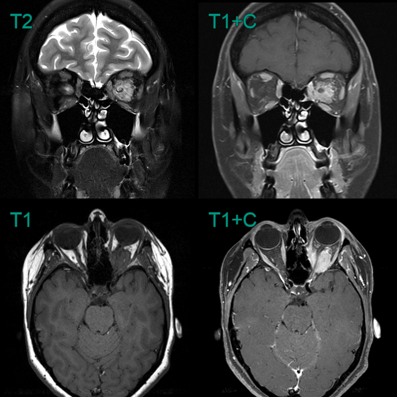
- 40-year-old patient presenting with left sided proptosis.
- MRI showed an avidly enhancing lesion filling the orbital apex and encasing the optic nerve.
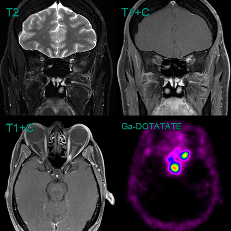
- 45-year-old female with eccentric enhancement of the left optic nerve sheath near the orbital apex.
- There was very high uptake on the Ga-DOTATATE PET scan.
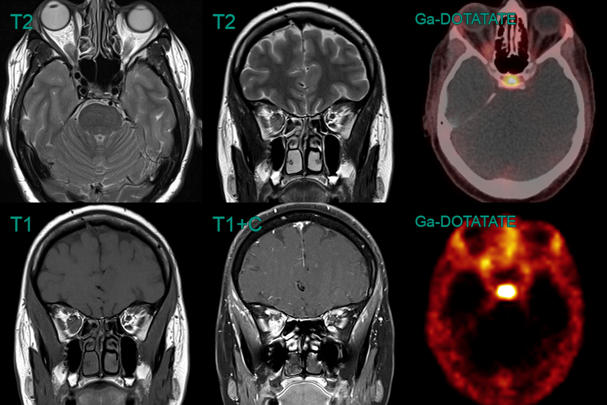
- 35-year-old patient developed worsening orbital swelling during pregnancy.
- An enhancing mass lesion along the inferior aspect of the optic nerve sheath, causing 2 mm of proptosis, was avid on Ga-DOTATATE PET.
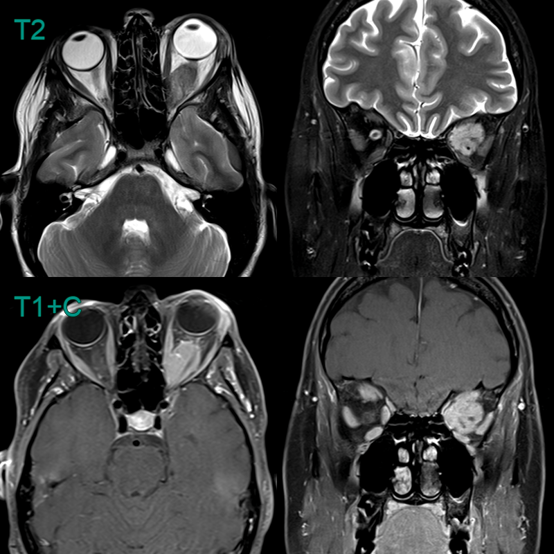
- 30-year-old patient presented with proptosis but no visual symptoms.
- MRI showed proptosis secondary to a large enhancing lesion encasing the optic nerve (red arrow).