Petrous cephalocoele
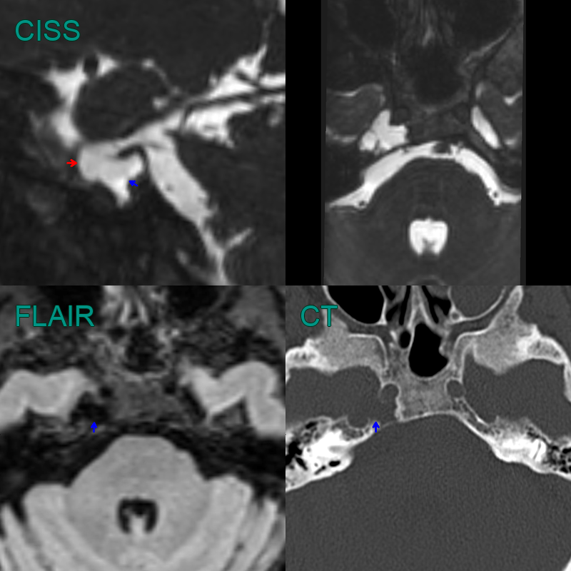
- Incidental finding of a CSF filled structure in the petrous apex (blue arrow).
- The cephalocoele was contiguous with Meckel's cave (red arrow).
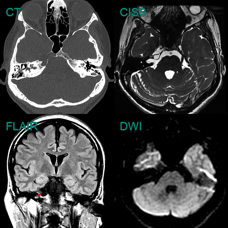
- A 50-year-old patient presentd with right sided trigeminal neuralgia.
- CT showed a well demarcated excavation of the right petrous apex.
- The cavity was filled with CSF with no enhancement of diffusion restriction.
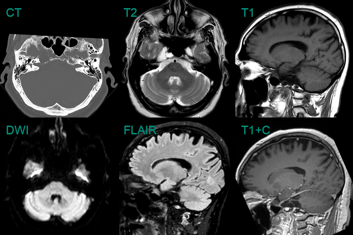
- A 60-year-old patient presented with headache.
- CT showed a well-marginated lesion remodelling the right petrous apex.
- MRI did not show any abnormal soft tissue or enhancement with only CSF signal content.