Primary CNS lymphoma
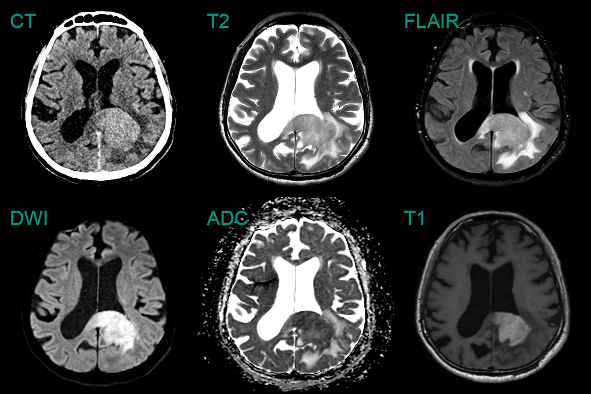
- 70-year-old patient presented with a 3 week history of headaches.
- CT showed a grossly enlarged and subtly hyperattenuating splenium.
- On MRI, the lesion enhanced homogeneously with diffusion restriction.
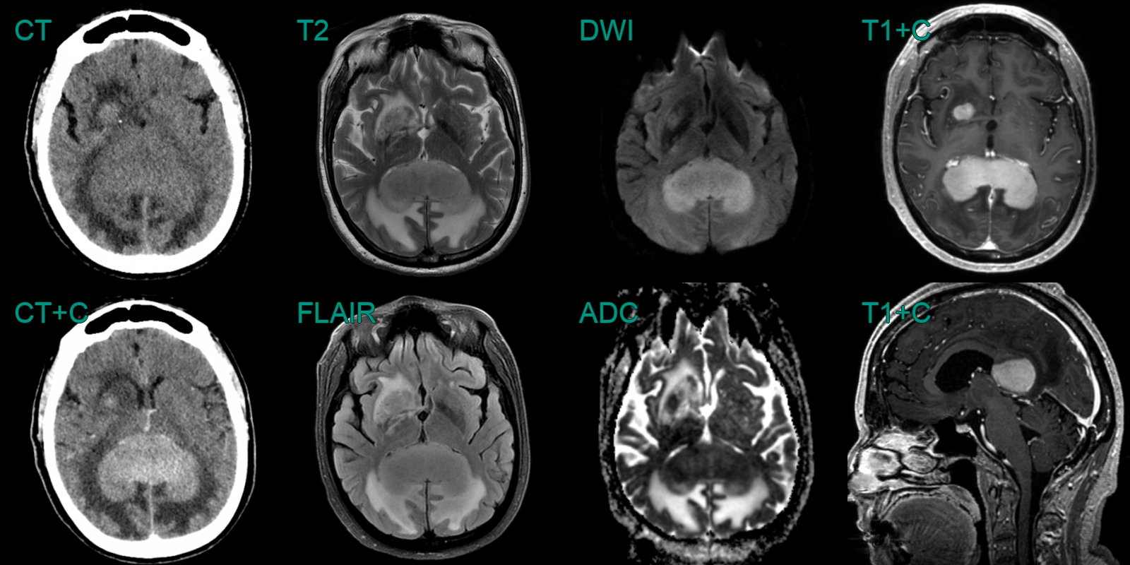
- A 70-year-old patient presented with confusion and visual disturbance.
- CT showed a hyperdense lesion involving the parietal white matter and corpus callosum.
- Relative hypointensity on T2 and low values on ADC also indicated hypercellularity.
- Alongside confluent avid enhancement, the imaging was typical for the final diagnosis of PCNSL.
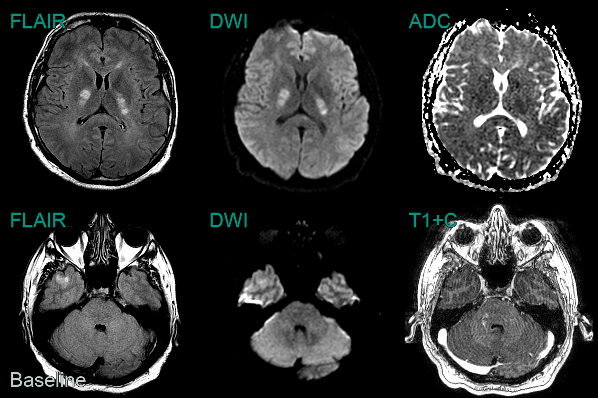
- A 40-year-old patient presented with agitation and aggressive behaviour.
- MRI showed mutliple lesions, some of which caused diffusion restriction (internal capsules) and some of which enhanced (right hemipons).
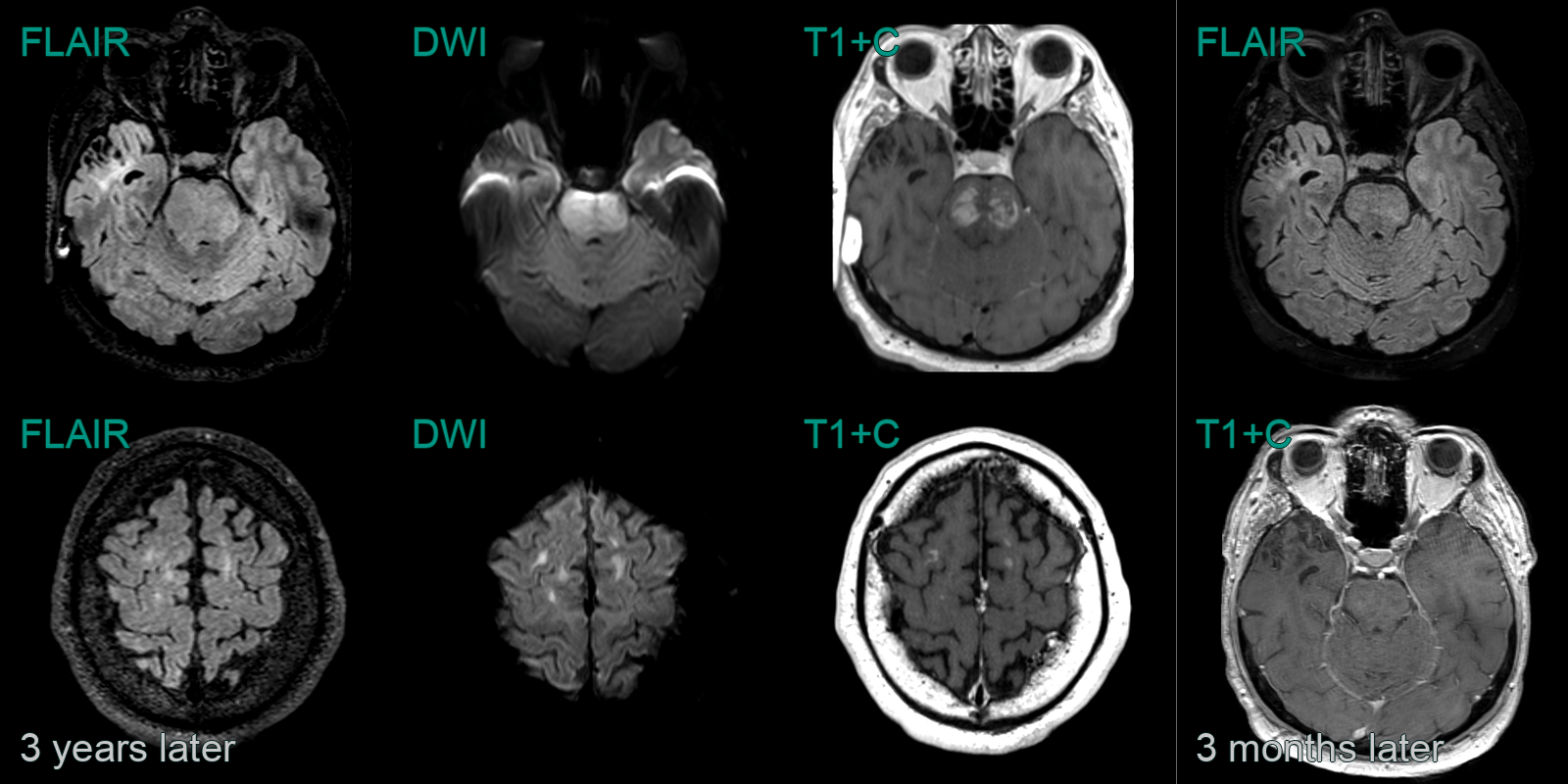
- A diagnosis was not secured even after extensive investigation. The lesions responded to a trial of steroid therapy.
- 3 years later, the patient presented following a seizure. MRI showed marked progression with an infiltative and enhancing lesion in the brainstem and new frontal lobe lesions.
- Biopsy of a right frontal lesion revealed a diffuse large B-cell lymphoma, which again showed a response to therapy.