Progressive Multifocal Leukoencephalopathy (PML)
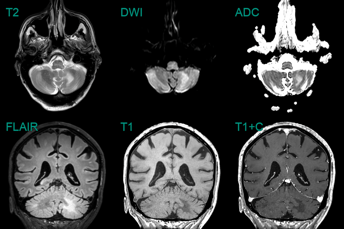
- 75-year-old patient on immunosuppression for rhematoid arthritis presented with subacute cerebellar ataxia.
- MRI showed a T2 and diffusion-weighted hyperintensity in the cerebellar white matter without enhancement.
- JC virus was positive and the lesions regressed after cessation of immunosuppressants.
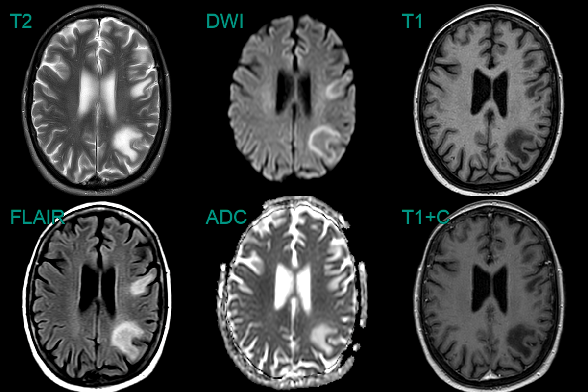
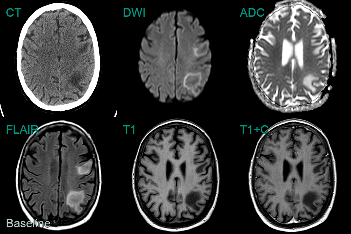
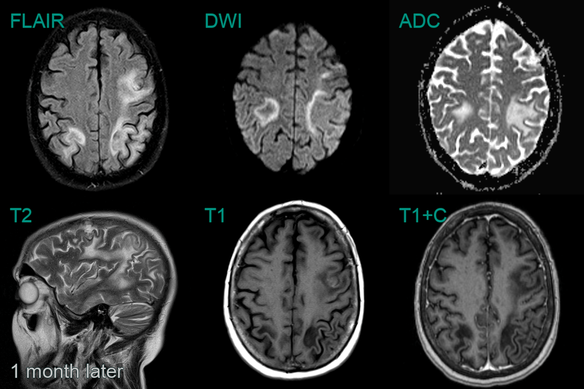
- 70-year-old patient undergoing treatment for lymphoma. Presented with seizures, confusion, and aphasia.
- MRI showed peripheral FLAIR-hyperintense and T1-hypointense lesions extending up to the cortex with no mass effect or enhancement.
- After one month and treatment with pembrolizumab, the lesions had enlarged with a more obvious leading edge of diffusion weighted hyperintensity. There was no contrast enhancement to suggest PML-IRIS.
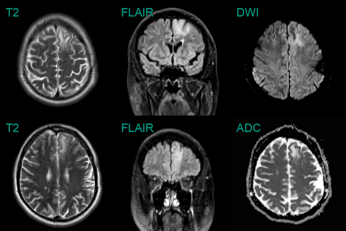
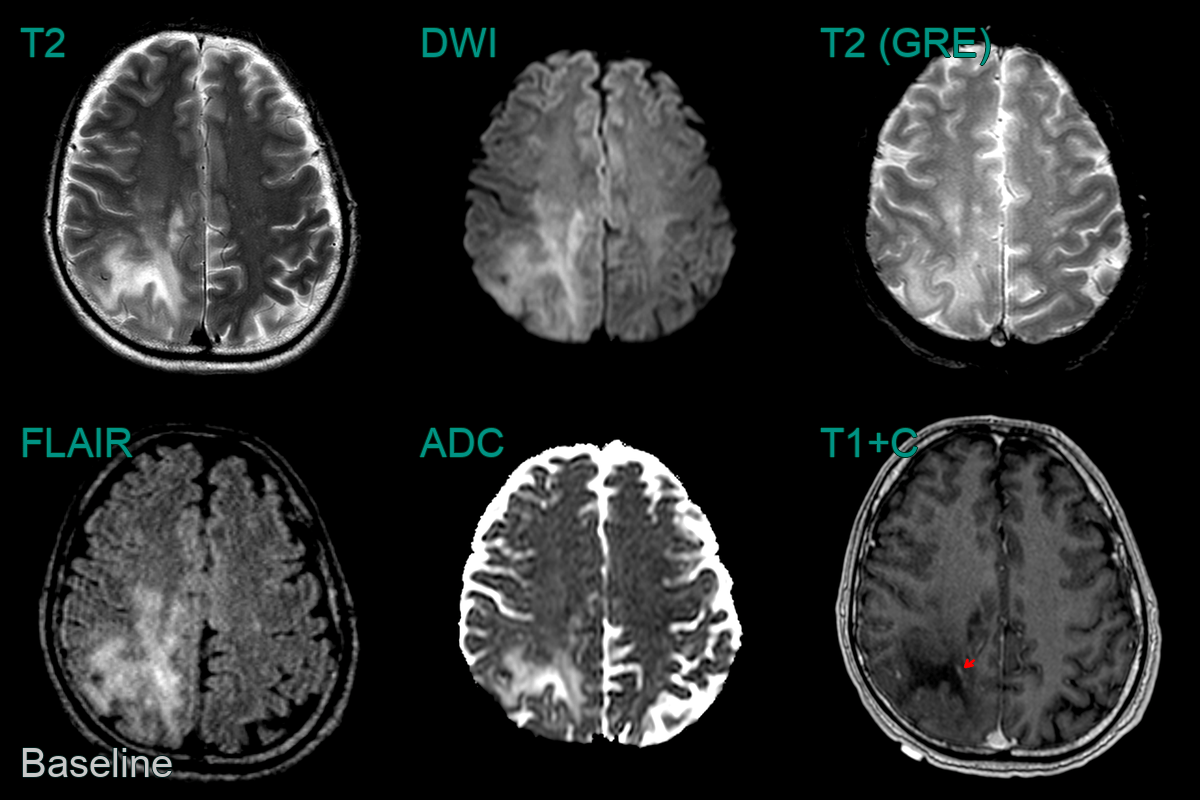
- A 40-year-old patient who had recently underwent CAR-T treatment for lymphoma presented after a 2 week history of headache and photophobia.
- MRI showed a large confluent subcortical region of T2-hyperintensity with a subtle rim of relative diffusion restriction and enhancement.
- Biopsy confirmed PML.
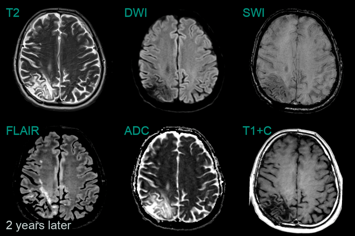
- On follow-up imaging 2 years later, following successful remission of lymphoma, the region matured into a region of gliosis.
