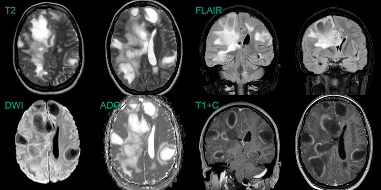
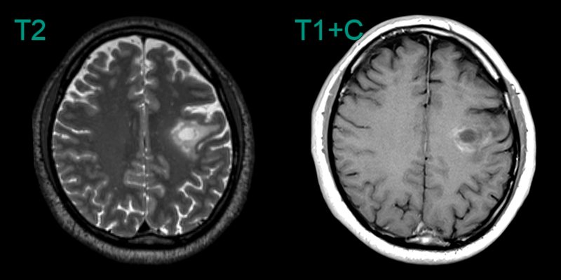
Tumefactive demyelination
- A 30-year-old patient presented with a left-sided facial droop and speech disturbance.
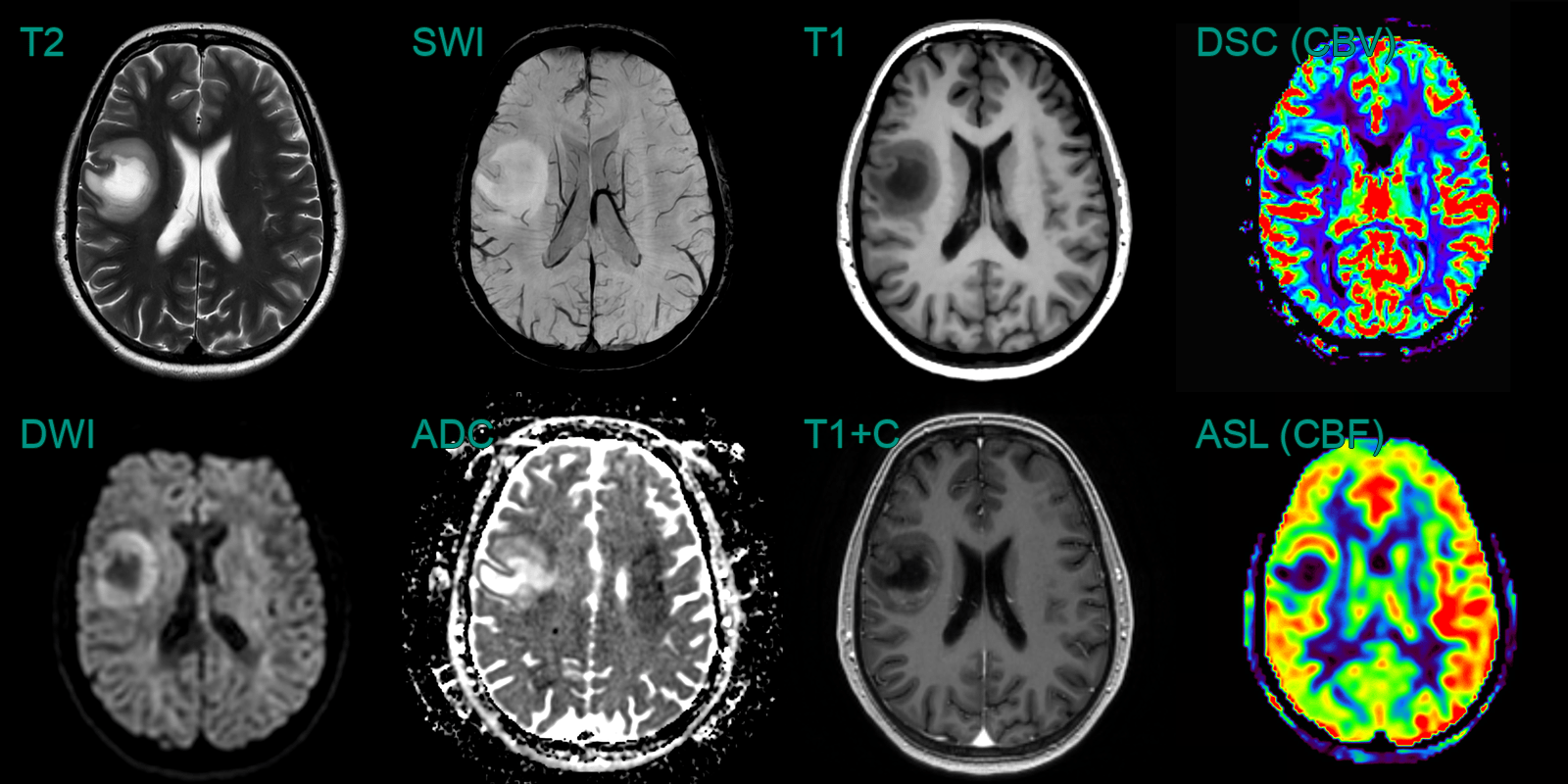
- MRI showed a T2-hyperintense lesion in the right posterior frontal lobe with a rim of enhancement and diffusion restriction.
- DSC and ASL perfusion showed elevated CBV and CBF peripherally (corresponding to the diffusion restriction).
- Biopsy confirmed inflammatory demyelination.


